Zaizen Yoshiaki, Tachibana Yuri, Kashima Yukio, Bychkov Andrey, Tabata Kazuhiro, Otani Kyoko, Kinoshita Yoshiaki, Yamano Yasuhiko, Kataoka Kensuke, Ichikado Kazuya, Okamoto Masaki, Kishaba Tomoo, Mito Remi, Nishimura Koichi, Yamasue Mari, Nabeshima Kazuki, Watanabe Kentaro, Kondoh Yasuhiro, Fukuoka Junya
Department of Pathology, Nagasaki University Graduate School of Biomedical Sciences, 1-7-1 Sakamoto, Nagasaki 852-8501, Japan.
Division of Respirology, Neurology and Rheumatology, Department of Medicine, Kurume University School of Medicine, 67 Asahi-machi, Kurume, Fukuoka 830-830-0011, Japan.
J Clin Med. 2021 Feb 24;10(5):895. doi: 10.3390/jcm10050895.
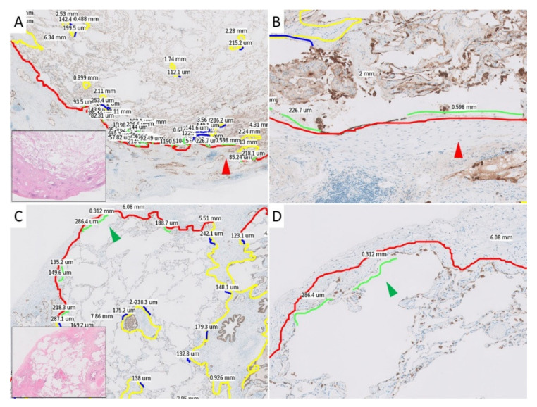
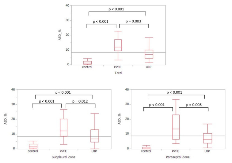
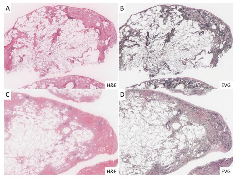
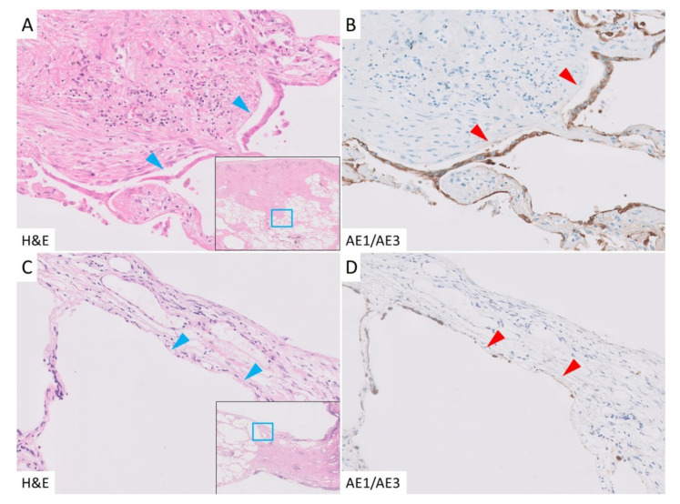
The pathogenesis of pleuroparenchymal fibroelastosis (PPFE), a rare interstitial lung disease, remains unclear. Based on previous reports and our experience, we hypothesized that alveolar epithelial denudation (AED) was involved in the pathogenesis of PPFE. This multicenter retrospective study investigated the percentage of AED and the features of the denudated areas in 26 PPFE cases, 30 idiopathic pulmonary fibrosis (IPF) cases, and 29 controls. PPFE patients had lower forced vital capacities and higher residual volume/total lung capacities in pulmonary function tests compared to IPF and control patients. Histopathologically, subpleural fibroelastosis was observed in PPFE, and AED was observed in 12.01% of cases in the subpleural or interlobular septa regardless of fibroelastosis. The percentage of AED in the PPFE group was significantly higher than that in the IPF group (6.84%; = 0.03) and the normal group (1.19%; < 0.001). In the IPF group, the percentage of AED and the presence of PPFE-like lesions in the upper lobes were examined radiologically, but no correlation was found. We showed that AED frequently occurred in PPFE. AED was less frequent in IPF, which, in combination with imaging data, suggests that PPFE may have a different pathogenesis from IPF.
胸膜实质纤维弹性组织增生症(PPFE)是一种罕见的间质性肺疾病,其发病机制尚不清楚。基于既往报道和我们的经验,我们推测肺泡上皮剥脱(AED)参与了PPFE的发病机制。这项多中心回顾性研究调查了26例PPFE患者、30例特发性肺纤维化(IPF)患者和29名对照者的AED百分比及剥脱区域的特征。与IPF患者和对照者相比,PPFE患者在肺功能测试中的用力肺活量较低,残气量/肺总量较高。组织病理学上,在PPFE中观察到胸膜下纤维弹性组织增生,无论有无纤维弹性组织增生,在胸膜下或小叶间隔中12.01%的病例中观察到AED。PPFE组的AED百分比显著高于IPF组(6.84%;P = 0.03)和正常组(1.19%;P < 0.001)。在IPF组中,通过影像学检查了AED百分比及上叶中PPFE样病变的存在情况,但未发现相关性。我们发现AED在PPFE中频繁发生。AED在IPF中较少见,结合影像学数据,提示PPFE可能具有与IPF不同的发病机制。