Ueno Harushi, Yazawa Mari, Tsubouchi Hideki, Nakanishi Keita, Sugiyama Tomoshi, Kadomatsu Yuka, Goto Masaki, Ozeki Naoki, Nakamura Shota, Fukui Takayuki, Mutsuga Masato, Chen Yoshikawa Toyofumi Fengshi
Department of Thoracic Surgery, Nagoya University Graduate School of Medicine, 65 Tsurumai-cho, Showa-ku, Nagoya, 466-8550, Japan.
Department of Cardiac Surgery, Nagoya University Graduate School of Medicine, 65 Tsurumai-cho, Showa-ku, Nagoya, 466-8550, Japan.
Surg Case Rep. 2021 Mar 9;7(1):66. doi: 10.1186/s40792-021-01148-0.
Aneurysm of the left brachiocephalic vein is a very rare clinical disease and only 40 cases have been reported so far.
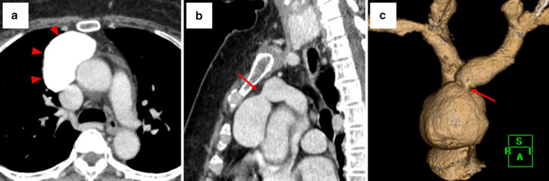
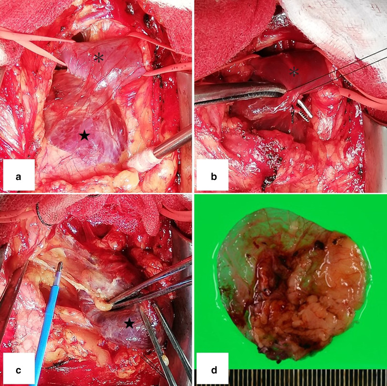
The patient was a 61-year-old woman with no related medical history. She underwent CT to investigate the cause of a cough and a mass was noted in the anterior mediastinum. Dynamic computed tomography with contrast medium injected into the left basilic vein demonstrated the venous aneurysm with blood flow to the left brachiocephalic vein. The patient had no symptoms, but because of the risk of pulmonary infarction and aneurysm rupture, the aneurysm was surgically resected. A median sternotomy was a reasonable approach because of the fragility of the venous aneurysm wall with little working space in the anterior mediastinum.
We diagnosed an aneurysm of the left brachiocephalic vein on preoperative imaging and excised it through a median sternotomy. The venous wall was thin and fragile in some areas and so this approach was appropriate in view of the possibility of intraoperative injury.
左头臂静脉动脉瘤是一种非常罕见的临床疾病,迄今为止仅报道了40例。
该患者为一名61岁女性,无相关病史。她接受了CT检查以调查咳嗽的原因,结果在前纵隔发现了一个肿块。向左侧贵要静脉注入造影剂的动态计算机断层扫描显示了静脉动脉瘤以及流向左头臂静脉的血流。患者没有症状,但由于存在肺梗死和动脉瘤破裂的风险,对动脉瘤进行了手术切除。由于静脉动脉瘤壁脆弱且前纵隔工作空间小,正中胸骨切开术是一种合理的方法。
我们在术前影像学检查中诊断出左头臂静脉动脉瘤,并通过正中胸骨切开术将其切除。静脉壁在某些区域薄且脆弱,因此鉴于术中受伤的可能性,这种方法是合适的。