Wegrzyn David, Freund Nadja, Faissner Andreas, Juckel Georg
Department of Cell Morphology and Molecular Neurobiology, Ruhr-University Bochum, Bochum, Germany.
Division of Experimental and Molecular Psychiatry, Department of Psychiatry, Psychotherapy and Preventive Medicine, LWL University Hospital, Ruhr-University Bochum, Bochum, Germany.
Front Synaptic Neurosci. 2021 Feb 23;13:637549. doi: 10.3389/fnsyn.2021.637549. eCollection 2021.
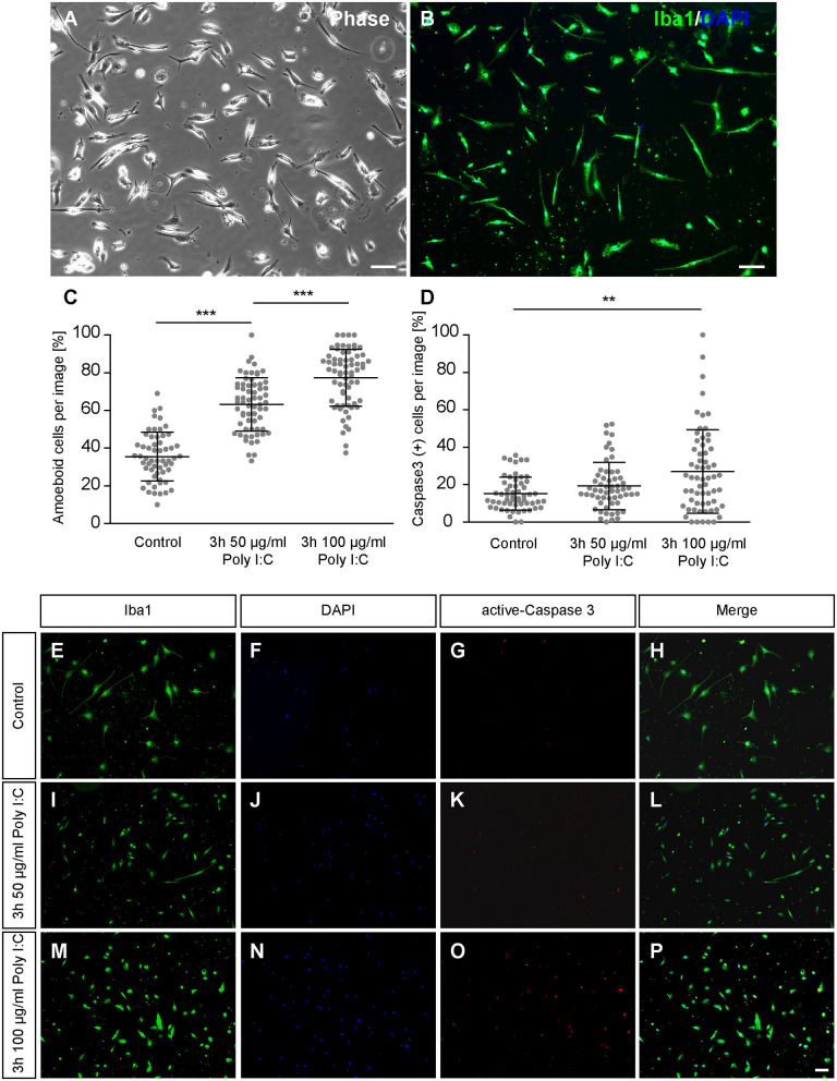
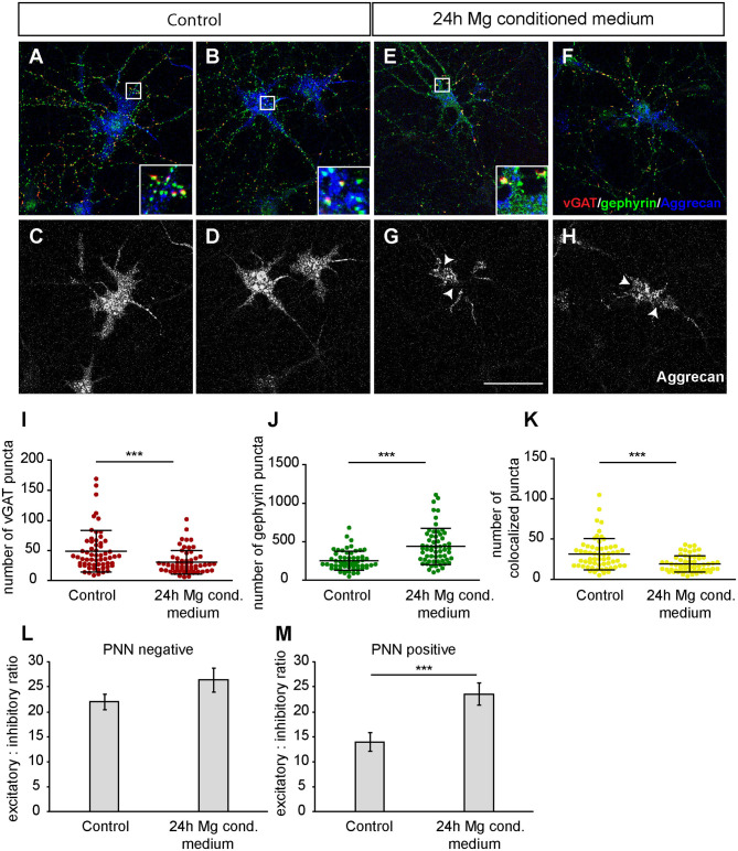
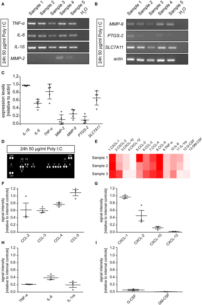
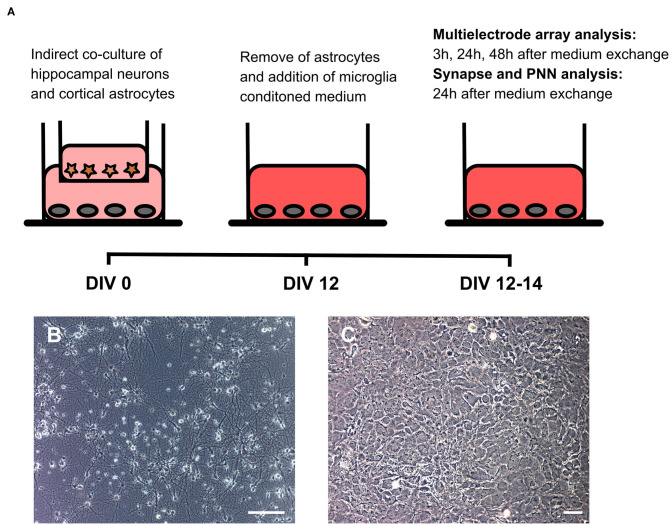
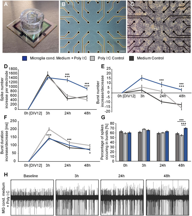
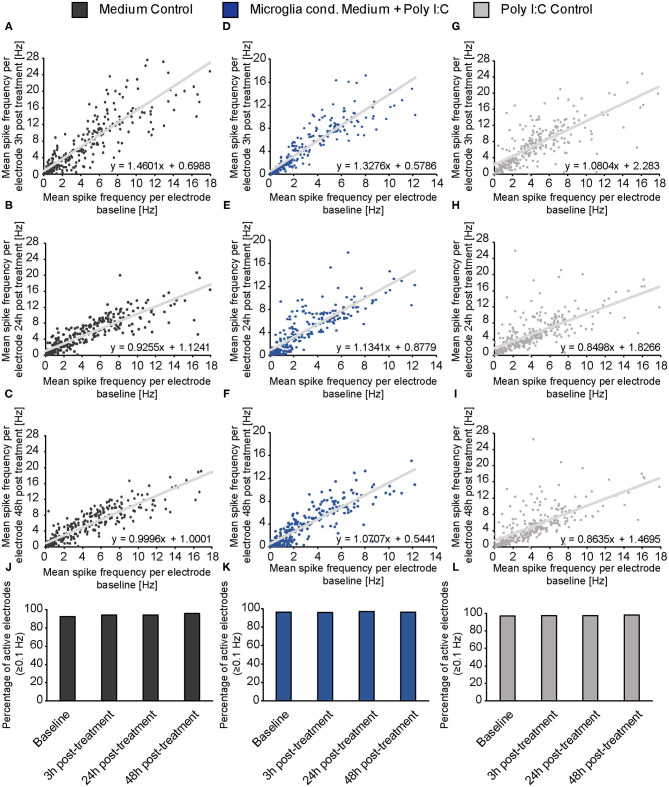
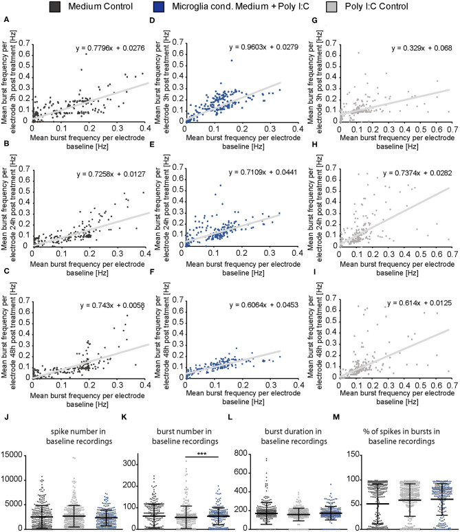
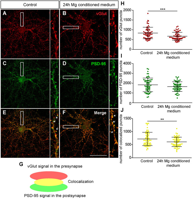
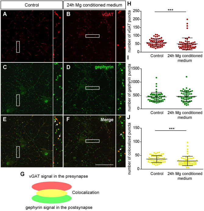
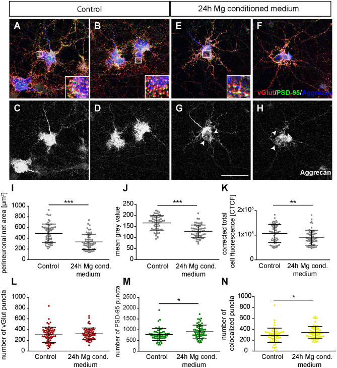
Perineuronal nets (PNNs) are specialized, reticular structures of the extracellular matrix (ECM) that can be found covering the soma and proximal dendrites of a neuronal subpopulation. Recent studies have shown that PNNs can highly influence synaptic plasticity and are disrupted in different neuropsychiatric disorders like schizophrenia. Interestingly, there is a growing evidence that microglia can promote the loss of PNNs and contribute to neuropsychiatric disorders. Based on this knowledge, we analyzed the impact of activated microglia on hippocampal neuronal networks . Therefore, primary cortical microglia were cultured and stimulated via polyinosinic-polycytidylic acid (Poly I:C; 50 μg/ml) administration. The Poly I:C treatment induced the expression and secretion of different cytokines belonging to the CCL- and CXCL-motif chemokine family as well as interleukin-6 (IL-6) and tumor necrosis factor-α (TNF-α). In addition, the expression of matrix metalloproteinases (MMPs) could be verified via RT-PCR analysis. Embryonic hippocampal neurons were then cultured for 12 days (DIV) and treated for 24 h with microglial conditioned medium. Interestingly, immunocytochemical staining of the PNN component Aggrecan revealed a clear disruption of PNNs accompanied by a significant increase of glutamatergic and a decrease of γ-aminobutyric acid-(GABA)ergic synapse numbers on PNN wearing neurons. In contrast, PNN negative neurons showed a significant reduction in both, glutamatergic and GABAergic synapses. Electrophysiological recordings were performed via multielectrode array (MEA) technology and unraveled a significantly increased spontaneous network activity that sustained also 24 and 48 h after the administration of microglia conditioned medium. Taken together, we could observe a strong impact of microglial secreted factors on PNN integrity, synaptic plasticity and electrophysiological properties of cultured neurons. Our observations might enhance the understanding of neuron-microglia interactions considering the ECM.
神经元周围网(PNNs)是细胞外基质(ECM)的特殊网状结构,可覆盖神经元亚群的胞体和近端树突。最近的研究表明,PNNs可对突触可塑性产生高度影响,并且在精神分裂症等不同神经精神疾病中会遭到破坏。有趣的是,越来越多的证据表明,小胶质细胞可促使PNNs丧失,并导致神经精神疾病。基于这一认识,我们分析了活化的小胶质细胞对海马神经元网络的影响。因此,培养原代皮质小胶质细胞,并通过给予多聚肌苷酸-多聚胞苷酸(Poly I:C;50μg/ml)进行刺激。Poly I:C处理诱导了属于CCL-和CXCL-基序趋化因子家族的不同细胞因子以及白细胞介素-6(IL-6)和肿瘤坏死因子-α(TNF-α)的表达和分泌。此外,可通过逆转录聚合酶链反应(RT-PCR)分析验证基质金属蛋白酶(MMPs)的表达。然后将胚胎海马神经元培养12天(培养天数,DIV),并用小胶质细胞条件培养基处理24小时。有趣的是,PNN成分聚集蛋白聚糖的免疫细胞化学染色显示PNNs明显遭到破坏,同时PNN包绕神经元上的谷氨酸能突触数量显著增加,γ-氨基丁酸(GABA)能突触数量减少。相比之下,PNN阴性神经元的谷氨酸能和GABA能突触均显著减少。通过多电极阵列(MEA)技术进行电生理记录,结果显示自发网络活动显著增加,在给予小胶质细胞条件培养基后24小时和48小时仍持续存在。综上所述,我们可以观察到小胶质细胞分泌因子对培养神经元的PNN完整性、突触可塑性和电生理特性有强烈影响。考虑到细胞外基质,我们的观察结果可能会增进对神经元-小胶质细胞相互作用的理解。