Matmusaev Maruf, Kumar R Senthil, Yamada Yasuhiro, Nagatani Tetsuya, Kawase Tsukasa, Tanaka Riki, Kyosuke Miyatani, Kato Yoko
Republican Specialized Scientific and Practical Medical Center of Neurosurgery, Tashkent, Uzbekistan.
Department of Neurosurgery, Royal Care Super Speciality Hospital, Coimbatore, Tamil Nadu, India.
Asian J Neurosurg. 2020 Oct 18;15(4):833-838. doi: 10.4103/ajns.AJNS_152_20. eCollection 2020 Oct-Dec.
Hemifacial spasm (HFS) is a condition, characterized by painless, involuntary unilateral tonic or clonic contractions of the facial muscles innervated by the ipsilateral facial nerve. HFS starts with contractions in the orbicularis oculi muscle with subsequent eyelid closure and/or eyebrow elevation, but may spread to involve muscles of the frontalis, platysma, and orbicularis oris muscles. Microvascular decompression (MVD) is reliable and accepted surgical treatment for HFS. MVD is the standard surgical technique now for HFS treatment with long-term success rates.
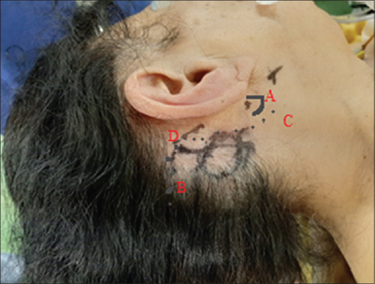
We performed fully endoscopic MVD technique for 1 patient with HFS (a 83-year-old female) at our institution. HFS was diagnosed based on the clinical history and presentation, a neurologic examination, and additional imaging findings. Respectively, the durations of HFS were 3 years, respectively. The patient had been previously treated with repeated botulinum toxin injections. Preoperative evaluation was done with magnetic resonance imaging; three-dimensional computed tomography fusion images examinations had identified the anterior inferior cerebellar artery (AICA) as the offending vessel in this patient.
The patient with HFS was treated by fully endoscopic MVD technique. The AICA, which had been identified as the offending vessel by preoperative magnetic resonance imaging, was successfully decompressed. No surgery-related complications occurred and had excellent outcomes with the complete resolution of HFS immediately after the operation.
Endoscopic surgery can provide a more panoramic surgical view than conventional microscopic surgery. Fully endoscopic MVD is both safe and effective in the treatment of HFS. This method minimizes the risks of brain retraction and extensive dissection often required for microscopic exposure. Endoscopic MVD is safe and has advantage over microscope in terms of visualization of structure, identification of neurovascular conflict, but it has a learning curve and technically challenging.
面肌痉挛(HFS)是一种以同侧面神经支配的面部肌肉无痛性、不自主单侧强直性或阵挛性收缩为特征的病症。HFS始于眼轮匝肌收缩,随后出现眼睑闭合和/或眉毛上抬,但可能蔓延至额肌、颈阔肌和口轮匝肌。微血管减压术(MVD)是治疗HFS可靠且被认可的手术方法。MVD是目前治疗HFS的标准手术技术,具有较高的长期成功率。
我们在本机构为1例HFS患者(一名83岁女性)实施了完全内镜下MVD技术。根据临床病史、表现、神经系统检查及其他影像学检查结果诊断为HFS。HFS的病程均为3年。该患者此前曾反复接受肉毒杆菌毒素注射治疗。术前通过磁共振成像进行评估;三维计算机断层扫描融合图像检查确定小脑前下动脉(AICA)为该患者的责任血管。
该HFS患者接受了完全内镜下MVD技术治疗。术前磁共振成像确定的责任血管AICA成功减压。未发生与手术相关的并发症,术后HFS立即完全缓解,效果极佳。
与传统显微手术相比,内镜手术可提供更全景的手术视野。完全内镜下MVD治疗HFS安全有效。该方法将显微暴露通常所需的脑牵拉和广泛解剖的风险降至最低。内镜下MVD安全,在结构可视化、神经血管冲突识别方面优于显微镜,但存在学习曲线且技术难度较大。