• 文献检索
  • 文档翻译
  • 深度研究
  • 学术资讯
  • Suppr Zotero 插件Zotero 插件
  • 邀请有礼
  • 套餐&价格
  • 历史记录
应用&插件
Suppr Zotero 插件Zotero 插件浏览器插件Mac 客户端Windows 客户端微信小程序
定价
高级版会员购买积分包购买API积分包
服务
文献检索文档翻译深度研究API 文档MCP 服务
关于我们
关于 Suppr公司介绍联系我们用户协议隐私条款
关注我们

Suppr 超能文献

核心技术专利:CN118964589B侵权必究
粤ICP备2023148730 号-1Suppr @ 2026

文献检索

告别复杂PubMed语法,用中文像聊天一样搜索,搜遍4000万医学文献。AI智能推荐,让科研检索更轻松。

立即免费搜索

文件翻译

保留排版,准确专业,支持PDF/Word/PPT等文件格式,支持 12+语言互译。

免费翻译文档

深度研究

AI帮你快速写综述,25分钟生成高质量综述,智能提取关键信息,辅助科研写作。

立即免费体验

细胞坏死性凋亡中鼠 RIPK3(受体相互作用蛋白激酶 3)的淀粉样结构。

The amyloid structure of mouse RIPK3 (receptor interacting protein kinase 3) in cell necroptosis.

机构信息

School of Life Science and Technology, ShanghaiTech University, Shanghai, PR China.

CAS Center for Excellence in Molecular Cell Science, Shanghai Institute of Biochemistry and Cell Biology, Chinese Academy of Sciences, Shanghai, PR China.

出版信息

Nat Commun. 2021 Mar 12;12(1):1627. doi: 10.1038/s41467-021-21881-2.

DOI:10.1038/s41467-021-21881-2
PMID:33712586
原文链接:https://pmc.ncbi.nlm.nih.gov/articles/PMC7955032/
Abstract

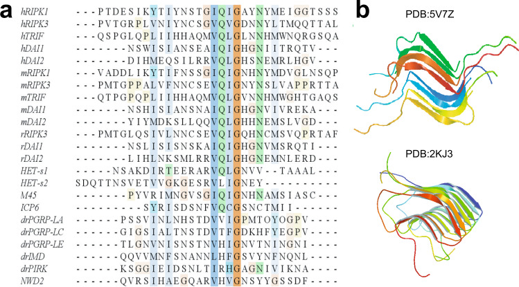
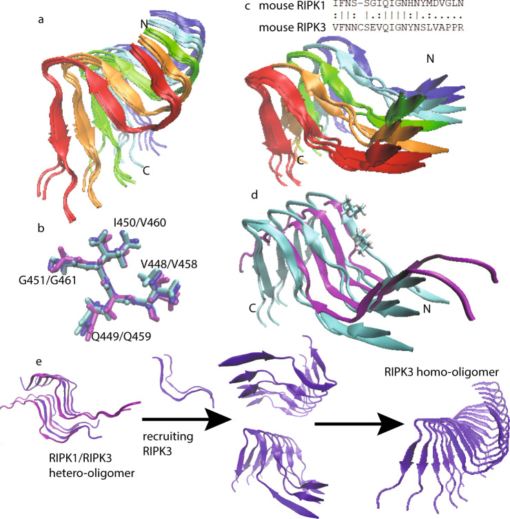
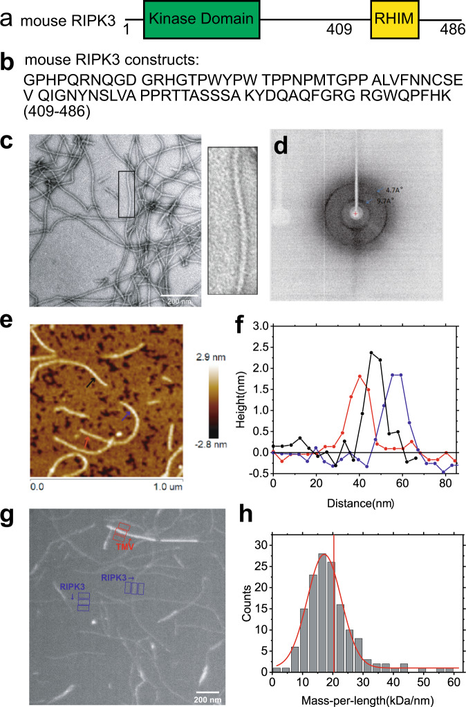
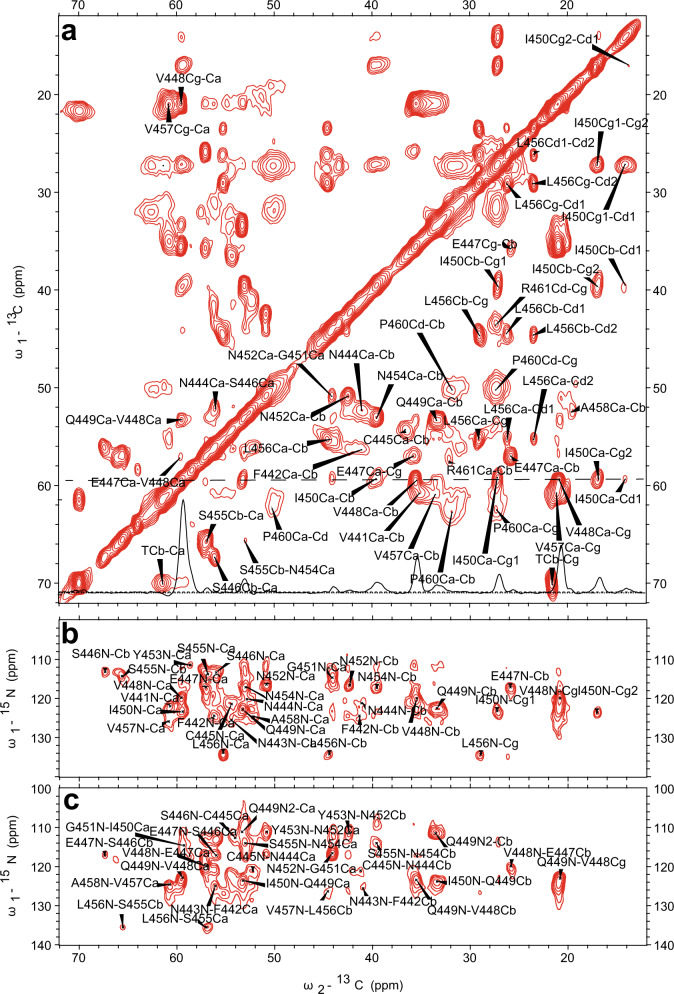
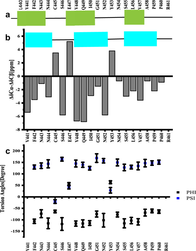
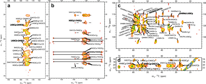
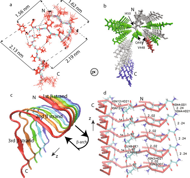
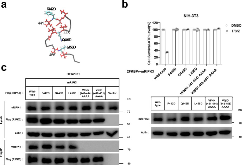
RIPK3 amyloid complex plays crucial roles during TNF-induced necroptosis and in response to immune defense in both human and mouse. Here, we have structurally characterized mouse RIPK3 homogeneous self-assembly using solid-state NMR, revealing a well-ordered N-shaped amyloid core structure featured with 3 parallel in-register β-sheets. This structure differs from previously published human RIPK1/RIPK3 hetero-amyloid complex structure, which adopted a serpentine fold. Functional studies indicate both RIPK1-RIPK3 binding and RIPK3 amyloid formation are essential but not sufficient for TNF-induced necroptosis. The structural integrity of RIPK3 fibril with three β-strands is necessary for signaling. Molecular dynamics simulations with a mouse RIPK1/RIPK3 model indicate that the hetero-amyloid is less stable when adopting the RIPK3 fibril conformation, suggesting a structural transformation of RIPK3 from RIPK1-RIPK3 binding to RIPK3 amyloid formation. This structural transformation would provide the missing link connecting RIPK1-RIPK3 binding to RIPK3 homo-oligomer formation in the signal transduction.

摘要

RIPK3 淀粉样复合物在 TNF 诱导的细胞坏死和人类及小鼠免疫防御中发挥关键作用。在此,我们使用固态 NMR 对小鼠 RIPK3 同型寡聚体进行了结构表征,揭示了一种有序的 N 型淀粉样核心结构,具有 3 个平行的顺序 β-折叠。该结构与先前发表的人源 RIPK1/RIPK3 异源淀粉样复合物结构不同,后者采用蛇形折叠。功能研究表明,RIPK1-RIPK3 结合和 RIPK3 淀粉样形成对于 TNF 诱导的细胞坏死是必需的,但不是充分的。具有三个 β-链的 RIPK3 纤维的结构完整性对于信号转导是必要的。使用小鼠 RIPK1/RIPK3 模型的分子动力学模拟表明,异源淀粉样采用 RIPK3 纤维构象时不太稳定,这表明 RIPK3 从 RIPK1-RIPK3 结合到 RIPK3 淀粉样形成的结构转换。这种结构转换将提供连接 RIPK1-RIPK3 结合到信号转导中 RIPK3 同源寡聚体形成的缺失环节。

https://cdn.ncbi.nlm.nih.gov/pmc/blobs/63f5/7955032/ee62538eb8b1/41467_2021_21881_Fig8_HTML.jpg
https://cdn.ncbi.nlm.nih.gov/pmc/blobs/63f5/7955032/0c56f9380106/41467_2021_21881_Fig1_HTML.jpg
https://cdn.ncbi.nlm.nih.gov/pmc/blobs/63f5/7955032/36d3fa597dfc/41467_2021_21881_Fig2_HTML.jpg
https://cdn.ncbi.nlm.nih.gov/pmc/blobs/63f5/7955032/8d857bcdc81e/41467_2021_21881_Fig3_HTML.jpg
https://cdn.ncbi.nlm.nih.gov/pmc/blobs/63f5/7955032/a434a4718294/41467_2021_21881_Fig4_HTML.jpg
https://cdn.ncbi.nlm.nih.gov/pmc/blobs/63f5/7955032/cc03af39472f/41467_2021_21881_Fig5_HTML.jpg
https://cdn.ncbi.nlm.nih.gov/pmc/blobs/63f5/7955032/ec118b20df0c/41467_2021_21881_Fig6_HTML.jpg
https://cdn.ncbi.nlm.nih.gov/pmc/blobs/63f5/7955032/ffb04ec01d7f/41467_2021_21881_Fig7_HTML.jpg
https://cdn.ncbi.nlm.nih.gov/pmc/blobs/63f5/7955032/ee62538eb8b1/41467_2021_21881_Fig8_HTML.jpg

相似文献

1
The amyloid structure of mouse RIPK3 (receptor interacting protein kinase 3) in cell necroptosis.细胞坏死性凋亡中鼠 RIPK3(受体相互作用蛋白激酶 3)的淀粉样结构。
Nat Commun. 2021 Mar 12;12(1):1627. doi: 10.1038/s41467-021-21881-2.
2
The structure of mouse RIPK1 RHIM-containing domain as a homo-amyloid and in RIPK1/RIPK3 complex.小鼠 RIPK1 RHIM 结构域作为同型淀粉样体和在 RIPK1/RIPK3 复合物中的结构。
Nat Commun. 2024 Aug 14;15(1):6975. doi: 10.1038/s41467-024-51303-y.
3
The Structure of the Necrosome RIPK1-RIPK3 Core, a Human Hetero-Amyloid Signaling Complex.坏死小体 RIPK1-RIPK3 核心结构,一种人类异源淀粉样信号复合物。
Cell. 2018 May 17;173(5):1244-1253.e10. doi: 10.1016/j.cell.2018.03.032. Epub 2018 Apr 19.
4
The structure of a minimum amyloid fibril core formed by necroptosis-mediating RHIM of human RIPK3.由人类 RIPK3 的 necroptosis 介导的 RHIM 形成的最小淀粉样原纤维核心的结构。
Proc Natl Acad Sci U S A. 2021 Apr 6;118(14). doi: 10.1073/pnas.2022933118.
5
FKBP12 mediates necroptosis by initiating RIPK1-RIPK3-MLKL signal transduction in response to TNF receptor 1 ligation.FKBP12 通过响应 TNF 受体 1 配体介导 RIPK1-RIPK3-MLKL 信号转导来介导坏死性凋亡。
J Cell Sci. 2019 May 20;132(10):jcs227777. doi: 10.1242/jcs.227777.
6
Human RIPK3 maintains MLKL in an inactive conformation prior to cell death by necroptosis.在细胞发生坏死性细胞死亡(necroptosis)之前,人源 RIPK3 将 MLKL 维持在非活性构象。
Nat Commun. 2021 Nov 22;12(1):6783. doi: 10.1038/s41467-021-27032-x.
7
Viral M45 and necroptosis-associated proteins form heteromeric amyloid assemblies.病毒 M45 和坏死性凋亡相关蛋白形成杂合的淀粉样组装体。
EMBO Rep. 2019 Feb;20(2). doi: 10.15252/embr.201846518. Epub 2018 Nov 29.
8
Identification of the Raf kinase inhibitor TAK-632 and its analogues as potent inhibitors of necroptosis by targeting RIPK1 and RIPK3.鉴定 Raf 激酶抑制剂 TAK-632 及其类似物为 RIPK1 和 RIPK3 的有效抑制剂,可抑制坏死性凋亡。
Br J Pharmacol. 2019 Jun;176(12):2095-2108. doi: 10.1111/bph.14653. Epub 2019 May 6.
9
RIPK1 inhibits ZBP1-driven necroptosis during development.RIPK1 抑制发育过程中 ZBP1 驱动的坏死性凋亡。
Nature. 2016 Dec 1;540(7631):129-133. doi: 10.1038/nature20559. Epub 2016 Nov 7.
10
Identification of MYC as an antinecroptotic protein that stifles RIPK1-RIPK3 complex formation.鉴定 MYC 作为一种抗细胞坏死蛋白,可抑制 RIPK1-RIPK3 复合物的形成。
Proc Natl Acad Sci U S A. 2020 Aug 18;117(33):19982-19993. doi: 10.1073/pnas.2000979117. Epub 2020 Aug 4.

引用本文的文献

1
Novel Delivery of Cyclic-Diguanylate Monophosphate Utilizing Amyloid Depots.利用淀粉样蛋白沉积物实现环二鸟苷单磷酸的新型递送。
Pharmaceutics. 2025 May 19;17(5):668. doi: 10.3390/pharmaceutics17050668.
2
RIPK1 is required for ZBP1-driven necroptosis in human cells.RIPK1是人类细胞中ZBP1驱动的坏死性凋亡所必需的。
PLoS Biol. 2025 Feb 21;23(2):e3002845. doi: 10.1371/journal.pbio.3002845. eCollection 2025 Feb.
3
Necroptosis in obesity: a complex cell death event.肥胖中的坏死性凋亡:一种复杂的细胞死亡事件。

本文引用的文献

1
RIP3-mediated necroptosis is regulated by inter-filament assembly of RIP homotypic interaction motif.RIP3 介导线粒体依赖性细胞坏死是通过 RIP 同源相互作用基序的细丝组装来调节的。
Cell Death Differ. 2021 Jan;28(1):251-266. doi: 10.1038/s41418-020-0598-9. Epub 2020 Jul 31.
2
The Killer Pseudokinase Mixed Lineage Kinase Domain-Like Protein (MLKL).杀手假激酶混合谱系激酶结构域样蛋白(MLKL)。
Cold Spring Harb Perspect Biol. 2020 Aug 3;12(8):a036376. doi: 10.1101/cshperspect.a036376.
3
A three-dimensional potential of mean force to improve backbone and sidechain hydrogen bond geometry in Xplor-NIH protein structure determination.
Apoptosis. 2025 Feb;30(1-2):466-487. doi: 10.1007/s10495-024-02055-z. Epub 2024 Dec 19.
4
Dipolar Recoupling in Rotating Solids.旋转固体中的偶极重耦合
Chem Rev. 2024 Nov 27;124(22):12844-12917. doi: 10.1021/acs.chemrev.4c00373. Epub 2024 Nov 6.
5
RIPK1 is essential for Herpes Simplex Virus-triggered ZBP1-dependent necroptosis in human cells.RIPK1对于单纯疱疹病毒引发的人类细胞中ZBP1依赖性坏死性凋亡至关重要。
bioRxiv. 2024 Sep 19:2024.09.17.613393. doi: 10.1101/2024.09.17.613393.
6
The structure of mouse RIPK1 RHIM-containing domain as a homo-amyloid and in RIPK1/RIPK3 complex.小鼠 RIPK1 RHIM 结构域作为同型淀粉样体和在 RIPK1/RIPK3 复合物中的结构。
Nat Commun. 2024 Aug 14;15(1):6975. doi: 10.1038/s41467-024-51303-y.
7
HSPA8 inhibitors augment cancer chemotherapeutic effectiveness via potentiating necroptosis.HSPA8 抑制剂通过增强细胞坏死作用增强癌症化疗疗效。
Mol Biol Cell. 2024 Aug 1;35(8):ar108. doi: 10.1091/mbc.E24-04-0194. Epub 2024 Jul 3.
8
Need for standardization of Influenza A virus-induced cell death in vivo to improve consistency of inter-laboratory research findings.需要对甲型流感病毒在体内诱导的细胞死亡进行标准化,以提高实验室间研究结果的一致性。
Cell Death Discov. 2024 May 22;10(1):247. doi: 10.1038/s41420-024-01981-w.
9
MLKL-mediated necroptosis is activated by RIPK1/3 and involved in anti-microbial immunity.MLKL 介导线粒体依赖性细胞坏死是由 RIPK1/3 激活的,并参与抗微生物免疫。
Front Immunol. 2024 Jan 16;15:1348866. doi: 10.3389/fimmu.2024.1348866. eCollection 2024.
10
RIP3 in Necroptosis: Underlying Contributions to Traumatic Brain Injury.RIP3 在细胞坏死性凋亡中的作用:对创伤性脑损伤的潜在贡献。
Neurochem Res. 2024 Feb;49(2):245-257. doi: 10.1007/s11064-023-04038-z. Epub 2023 Sep 25.
一种三维平均力势方法,用于改善 Xplor-NIH 蛋白质结构测定中主链和侧链氢键的几何形状。
Protein Sci. 2020 Jan;29(1):100-110. doi: 10.1002/pro.3745. Epub 2019 Oct 27.
4
ZBP1/DAI Drives RIPK3-Mediated Cell Death Induced by IFNs in the Absence of RIPK1.ZBP1/DAI 驱动 IFNs 诱导的 RIPK3 介导的细胞死亡,而无需 RIPK1。
J Immunol. 2019 Sep 1;203(5):1348-1355. doi: 10.4049/jimmunol.1900216. Epub 2019 Jul 29.
5
The PSIPRED Protein Analysis Workbench: 20 years on.PSIPRED 蛋白质分析工作平台:20 年的发展
Nucleic Acids Res. 2019 Jul 2;47(W1):W402-W407. doi: 10.1093/nar/gkz297.
6
Functional Amyloids.功能性淀粉样蛋白
Cold Spring Harb Perspect Biol. 2019 Dec 2;11(12):a033860. doi: 10.1101/cshperspect.a033860.
7
Viral M45 and necroptosis-associated proteins form heteromeric amyloid assemblies.病毒 M45 和坏死性凋亡相关蛋白形成杂合的淀粉样组装体。
EMBO Rep. 2019 Feb;20(2). doi: 10.15252/embr.201846518. Epub 2018 Nov 29.
8
The Structure of the Necrosome RIPK1-RIPK3 Core, a Human Hetero-Amyloid Signaling Complex.坏死小体 RIPK1-RIPK3 核心结构,一种人类异源淀粉样信号复合物。
Cell. 2018 May 17;173(5):1244-1253.e10. doi: 10.1016/j.cell.2018.03.032. Epub 2018 Apr 19.
9
Death-domain dimerization-mediated activation of RIPK1 controls necroptosis and RIPK1-dependent apoptosis.死亡结构域二聚化介导的 RIPK1 激活控制坏死性凋亡和 RIPK1 依赖性细胞凋亡。
Proc Natl Acad Sci U S A. 2018 Feb 27;115(9):E2001-E2009. doi: 10.1073/pnas.1722013115. Epub 2018 Feb 12.
10
Structure of FUS Protein Fibrils and Its Relevance to Self-Assembly and Phase Separation of Low-Complexity Domains.FUS蛋白原纤维的结构及其与低复杂性结构域的自组装和相分离的相关性。
Cell. 2017 Oct 19;171(3):615-627.e16. doi: 10.1016/j.cell.2017.08.048. Epub 2017 Sep 21.