• 文献检索
  • 文档翻译
  • 深度研究
  • 学术资讯
  • Suppr Zotero 插件Zotero 插件
  • 邀请有礼
  • 套餐&价格
  • 历史记录
应用&插件
Suppr Zotero 插件Zotero 插件浏览器插件Mac 客户端Windows 客户端微信小程序
定价
高级版会员购买积分包购买API积分包
服务
文献检索文档翻译深度研究API 文档MCP 服务
关于我们
关于 Suppr公司介绍联系我们用户协议隐私条款
关注我们

Suppr 超能文献

核心技术专利:CN118964589B侵权必究
粤ICP备2023148730 号-1Suppr @ 2026

文献检索

告别复杂PubMed语法,用中文像聊天一样搜索,搜遍4000万医学文献。AI智能推荐,让科研检索更轻松。

立即免费搜索

文件翻译

保留排版,准确专业,支持PDF/Word/PPT等文件格式,支持 12+语言互译。

免费翻译文档

深度研究

AI帮你快速写综述,25分钟生成高质量综述,智能提取关键信息,辅助科研写作。

立即免费体验

F-PSMA-1007 PET/CT、全身MRI和CT在不良中高危前列腺癌原发淋巴结分期中的前瞻性比较

Prospective comparison of F-PSMA-1007 PET/CT, whole-body MRI and CT in primary nodal staging of unfavourable intermediate- and high-risk prostate cancer.

作者信息

Malaspina Simona, Anttinen Mikael, Taimen Pekka, Jambor Ivan, Sandell Minna, Rinta-Kiikka Irina, Kajander Sami, Schildt Jukka, Saukko Ekaterina, Noponen Tommi, Saunavaara Jani, Dean Peter B, Sequeiros Roberto Blanco, Aronen Hannu J, Kemppainen Jukka, Seppänen Marko, Boström Peter J, Ettala Otto

机构信息

Turku PET Centre, University of Turku and Turku University Hospital, Turku, Finland.

Department of Urology, University of Turku and Turku University Hospital, Turku, Finland.

出版信息

Eur J Nucl Med Mol Imaging. 2021 Aug;48(9):2951-2959. doi: 10.1007/s00259-021-05296-1. Epub 2021 Mar 13.

DOI:10.1007/s00259-021-05296-1
PMID:33715033
原文链接:https://pmc.ncbi.nlm.nih.gov/articles/PMC8263440/
Abstract

PURPOSE

To prospectively compare F-prostate-specific membrane antigen (PSMA)-1007 positron emission tomography (PET)/CT, whole-body magnetic resonance imaging (WBMRI) including diffusion-weighted imaging (DWI) and standard computed tomography (CT), in primary nodal staging of prostate cancer (PCa).

METHODS

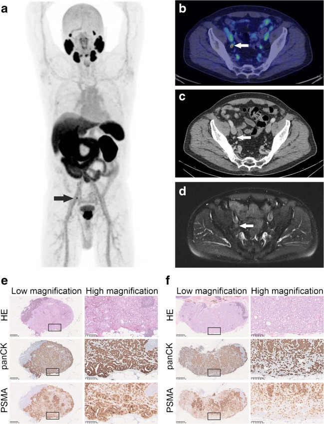
Men with newly diagnosed unfavourable intermediate- or high-risk PCa prospectively underwent F-PSMA-1007 PET/CT, WBMRI with DWI and contrast-enhanced CT within a median of 8 days. Six readers (two for each modality) independently reported pelvic lymph nodes as malignant, equivocal or benign while blinded to the other imaging modalities. Sensitivity, specificity and accuracy were reported according to optimistic (equivocal lesions interpreted as benign) and pessimistic (equivocal lesions interpreted as malignant) analyses. The reference standard diagnosis was based on multidisciplinary consensus meetings where available histopathology, clinical and follow-up data were used.

RESULTS

Seventy-nine patients completed all the imaging modalities, except for one case of interrupted WBMRI. Thirty-one (39%) patients had pelvic lymph node metastases, which were detected in 27/31 (87%), 14/31 (45%) and 8/31 (26%) patients by F-PSMA-1007 PET/CT, WBMRI with DWI and CT, respectively (optimistic analysis). In 8/31 (26%) patients, only F-PSMA-1007 PET/CT detected malignant lymph nodes, while the other two imaging modalities were reported as negative. At the patient level, sensitivity and specificity values for F-PSMA-1007 PET/CT, WBMRI with DWI and CT in optimistic analysis were 0.87 (95%CI 0.71-0.95) and 0.98 (95%CI 0.89-1.00), 0.37 (95%CI 0.22-0.55) and 0.98 (95%CI 0.89-1.00) and 0.26 (95%CI 0.14-0.43) and 1.00 (95%CI 0.93-1.00), respectively.

CONCLUSION

F-PSMA-1007 PET/CT showed significantly greater sensitivity in nodal staging of primary PCa than did WBMRI with DWI or CT, while maintaining high specificity.

CLINICAL TRIAL REGISTRATION

Clinicaltrials.gov ID: NCT03537391.

摘要

目的

前瞻性比较F-前列腺特异性膜抗原(PSMA)-1007正电子发射断层扫描(PET)/CT、包括扩散加权成像(DWI)的全身磁共振成像(WBMRI)和标准计算机断层扫描(CT)在前列腺癌(PCa)原发淋巴结分期中的应用。

方法

新诊断为不良中危或高危PCa的男性患者在中位8天内前瞻性地接受了F-PSMA-1007 PET/CT、带DWI的WBMRI和增强CT检查。六位阅片者(每种检查方式两位)在对其他成像方式不知情的情况下,独立将盆腔淋巴结报告为恶性、可疑或良性。根据乐观分析(将可疑病变解释为良性)和悲观分析(将可疑病变解释为恶性)报告敏感性、特异性和准确性。参考标准诊断基于多学科共识会议,会上使用了可用的组织病理学、临床和随访数据。

结果

79例患者完成了所有成像检查,除1例WBMRI检查中断。31例(39%)患者有盆腔淋巴结转移,F-PSMA-1007 PET/CT、带DWI的WBMRI和CT分别在27/31(87%)、14/31(45%)和8/31(26%)的患者中检测到转移(乐观分析)。在8/31(26%)的患者中,仅F-PSMA-1007 PET/CT检测到恶性淋巴结,而其他两种成像方式报告为阴性。在患者层面,乐观分析中F-PSMA-1007 PET/CT、带DWI的WBMRI和CT的敏感性和特异性值分别为0.87(95%CI 0.71 - 0.95)和0.98(95%CI 0.89 - 1.00)、0.37(95%CI 0.22 - 0.55)和0.98(95%CI 0.89 - 1.00)以及0.26(95%CI 0.14 - 0.43)和1.00(95%CI 0.93 - 1.00)。

结论

F-PSMA-1007 PET/CT在原发性PCa的淋巴结分期中显示出比带DWI的WBMRI或CT显著更高的敏感性,同时保持高特异性。

临床试验注册

Clinicaltrials.gov标识符:NCT03537391。

https://cdn.ncbi.nlm.nih.gov/pmc/blobs/f153/8263440/c375c506b669/259_2021_5296_Fig1_HTML.jpg

相似文献

1
Prospective comparison of F-PSMA-1007 PET/CT, whole-body MRI and CT in primary nodal staging of unfavourable intermediate- and high-risk prostate cancer.F-PSMA-1007 PET/CT、全身MRI和CT在不良中高危前列腺癌原发淋巴结分期中的前瞻性比较
Eur J Nucl Med Mol Imaging. 2021 Aug;48(9):2951-2959. doi: 10.1007/s00259-021-05296-1. Epub 2021 Mar 13.
2
A Prospective Comparison of F-prostate-specific Membrane Antigen-1007 Positron Emission Tomography Computed Tomography, Whole-body 1.5 T Magnetic Resonance Imaging with Diffusion-weighted Imaging, and Single-photon Emission Computed Tomography/Computed Tomography with Traditional Imaging in Primary Distant Metastasis Staging of Prostate Cancer (PROSTAGE).F-前列腺特异性膜抗原-1007 正电子发射断层扫描计算机断层扫描、全身 1.5T 磁共振成像弥散加权成像与传统成像单光子发射计算机断层扫描/计算机断层扫描在前列腺癌原发远处转移分期(PROSTAGE)中的前瞻性比较
Eur Urol Oncol. 2021 Aug;4(4):635-644. doi: 10.1016/j.euo.2020.06.012. Epub 2020 Jul 13.
3
More advantages in detecting bone and soft tissue metastases from prostate cancer using F-PSMA PET/CT.使用F-PSMA PET/CT检测前列腺癌骨和软组织转移方面有更多优势。
Hell J Nucl Med. 2019 Jan-Apr;22(1):6-9. doi: 10.1967/s002449910952. Epub 2019 Mar 7.
4
Lymph node staging with fluorine-18 prostate specific membrane antigen 1007-positron emission tomography/computed tomography in newly diagnosed intermediate- to high-risk prostate cancer using histopathological evaluation of extended pelvic node dissection as reference.使用经病理组织学评估的扩展盆腔淋巴结清扫术作为参考,对新诊断的中高危前列腺癌患者进行氟-18 前列腺特异性膜抗原 1007-正电子发射断层扫描/计算机断层扫描进行淋巴结分期。
Eur J Nucl Med Mol Imaging. 2022 Sep;49(11):3929-3937. doi: 10.1007/s00259-022-05827-4. Epub 2022 May 11.
5
Diagnostic Accuracy of F-PSMA-1007 PET/CT Imaging for Lymph Node Staging of Prostate Carcinoma in Primary and Biochemical Recurrence.F-PSMA-1007 PET/CT 成像在原发性和生化复发前列腺癌淋巴结分期中的诊断准确性。
J Nucl Med. 2021 Feb;62(2):208-213. doi: 10.2967/jnumed.120.246363. Epub 2020 Aug 17.
6
How accurate is Gallium-prostate specific membrane antigen positron emission tomography / computed tomography (Ga-PSMA PET/CT) on primary lymph node staging before radical prostatectomy in intermediate and high risk prostate cancer? A study of patient- and lymph node- based analyses.镓-前列腺特异性膜抗原正电子发射断层扫描/计算机断层扫描(Ga-PSMA PET/CT)在中高危前列腺癌根治性前列腺切除术前对原发性淋巴结分期的准确性如何?一项基于患者和淋巴结的分析研究。
Urol Oncol. 2022 Jan;40(1):6.e1-6.e9. doi: 10.1016/j.urolonc.2021.07.006. Epub 2021 Aug 13.
7
In primary lymph nodal staging of patients with high-risk and intermediate-risk prostate cancer, how critical is the role of Gallium-68 prostate-specific membrane antigen positron emission tomography-computed tomography?在高危和中危前列腺癌患者的原发性淋巴结分期中,镓68前列腺特异性膜抗原正电子发射断层扫描-计算机断层扫描的作用有多关键?
Nucl Med Commun. 2020 Feb;41(2):139-146. doi: 10.1097/MNM.0000000000001110.
8
Pelvic lymph-node staging with F-DCFPyL PET/CT prior to extended pelvic lymph-node dissection in primary prostate cancer - the SALT trial.在原发性前列腺癌中,使用 F-DCFPyL PET/CT 进行盆腔淋巴结分期后行扩大盆腔淋巴结清扫术 - SALT 试验。
Eur J Nucl Med Mol Imaging. 2021 Feb;48(2):509-520. doi: 10.1007/s00259-020-04974-w. Epub 2020 Aug 12.
9
Prostate-specific Membrane Antigen Positron Emission Tomography/Computed Tomography Compared with Conventional Imaging for Initial Staging of Treatment-naïve Intermediate- and High-risk Prostate Cancer: A Retrospective Single-center Study.前列腺特异性膜抗原正电子发射断层扫描/计算机断层扫描与常规成像在初治中高危前列腺癌分期中的比较:一项回顾性单中心研究。
Eur Urol Oncol. 2022 Oct;5(5):544-552. doi: 10.1016/j.euo.2020.08.012. Epub 2020 Sep 18.
10
Incremental Impact of [ Ga]Ga-PSMA-11 PET/CT in Primary N and M Staging of Prostate Cancer Prior to Curative-Intent Surgery: a Prospective Clinical Trial in Comparison with mpMRI.[68Ga]Ga-PSMA-11 PET/CT 对前列腺癌根治性手术前 N 和 M 分期的增量影响:与 mpMRI 比较的前瞻性临床试验。
Mol Imaging Biol. 2022 Feb;24(1):50-59. doi: 10.1007/s11307-021-01650-9. Epub 2021 Sep 14.

引用本文的文献

1
PSMA PET/CT for prostate cancer diagnosis: current applications and future directions.用于前列腺癌诊断的PSMA PET/CT:当前应用及未来方向
J Cancer Res Clin Oncol. 2025 May 4;151(5):155. doi: 10.1007/s00432-025-06184-z.
2
Prospective comparison of F-PSMA-1007 PET/CT and MRI with histopathology as the reference standard for intraprostatic tumour detection and T-staging of high-risk prostate cancer.以组织病理学为参考标准,对F-PSMA-1007 PET/CT和MRI在前列腺内肿瘤检测及高危前列腺癌T分期中的应用进行前瞻性比较。
Eur J Nucl Med Mol Imaging. 2025 Mar 31. doi: 10.1007/s00259-025-07208-z.
3
Comparative Performance of Ga-PSMA-11 PET/CT and Conventional Imaging in the Primary Staging of High-Risk Prostate Cancer Patients Who Are Candidates for Radical Prostatectomy.

本文引用的文献

1
Matched-Pair Comparison of Ga-PSMA-11 and F-rhPSMA-7 PET/CT in Patients with Primary and Biochemical Recurrence of Prostate Cancer: Frequency of Non-Tumor-Related Uptake and Tumor Positivity.镓-PSMA-11 和 F-rhPSMA-7 PET/CT 在初发和生化复发前列腺癌患者中的配对比较:非肿瘤相关摄取和肿瘤阳性的频率。
J Nucl Med. 2021 Aug 1;62(8):1082-1088. doi: 10.2967/jnumed.120.251447. Epub 2020 Dec 4.
2
Diagnostic Accuracy of F-PSMA-1007 PET/CT Imaging for Lymph Node Staging of Prostate Carcinoma in Primary and Biochemical Recurrence.F-PSMA-1007 PET/CT 成像在原发性和生化复发前列腺癌淋巴结分期中的诊断准确性。
J Nucl Med. 2021 Feb;62(2):208-213. doi: 10.2967/jnumed.120.246363. Epub 2020 Aug 17.
3
镓-PSMA-11 PET/CT与传统成像在适合根治性前列腺切除术的高危前列腺癌患者初始分期中的比较性能
Diagnostics (Basel). 2024 Sep 5;14(17):1964. doi: 10.3390/diagnostics14171964.
4
Multiparametric Whole-Body MRI: A Game Changer in Metastatic Prostate Cancer.多参数全身磁共振成像:转移性前列腺癌治疗的变革者
Cancers (Basel). 2024 Jul 13;16(14):2531. doi: 10.3390/cancers16142531.
5
Diagnostic Accuracy of Molecular Imaging Techniques for Detecting Prostate Cancer: A Systematic Review.分子成像技术检测前列腺癌的诊断准确性:一项系统评价。
Diagnostics (Basel). 2024 Jun 21;14(13):1315. doi: 10.3390/diagnostics14131315.
6
PSMA PET/CT in the low- to intermediate-risk prostate cancer: when and why?低至中危前列腺癌中的PSMA PET/CT:何时以及为何使用?
Asian J Androl. 2024 Nov 1;26(6):584-586. doi: 10.4103/aja20244. Epub 2024 Mar 26.
7
Preoperative evaluation of prostate cancer by Ga-PMSA positron emission tomography/computed tomography: comparison with magnetic resonance imaging and with histopathological findings.通过镓-前列腺特异性膜抗原正电子发射断层扫描/计算机断层扫描对前列腺癌进行术前评估:与磁共振成像及组织病理学结果的比较
Radiol Bras. 2023 Jul-Aug;56(4):171-178. doi: 10.1590/0100-3984.2022.0122-en.
8
18 F-prostate specific membrane antigen positron emission tomography/computerized tomography for lymph node staging in medium/high risk prostate cancer: A systematic review and meta-analysis.18F-前列腺特异性膜抗原正电子发射断层扫描/计算机断层扫描用于中/高危前列腺癌淋巴结分期:一项系统评价和荟萃分析
Chin Med J (Engl). 2024 Apr 20;137(8):949-958. doi: 10.1097/CM9.0000000000002850. Epub 2023 Sep 8.
9
[Research progress on prostate-specific membrane antigen ligand positron emission tomography imaging of prostate cancer].[前列腺癌前列腺特异性膜抗原配体正电子发射断层显像的研究进展]
Sheng Wu Yi Xue Gong Cheng Xue Za Zhi. 2022 Dec 25;39(6):1263-1268. doi: 10.7507/1001-5515.202112035.
10
Detection rate of fluorine-18 prostate-specific membrane antigen-1007 PET/CT for prostate cancer in primary staging and biochemical recurrence with different serum PSA levels: A systematic review and meta-analysis.氟-18前列腺特异性膜抗原-1007 PET/CT在不同血清前列腺特异抗原水平的前列腺癌原发分期及生化复发中的检测率:一项系统评价和Meta分析
Front Oncol. 2022 Jul 22;12:911146. doi: 10.3389/fonc.2022.911146. eCollection 2022.
Pelvic lymph-node staging with F-DCFPyL PET/CT prior to extended pelvic lymph-node dissection in primary prostate cancer - the SALT trial.
在原发性前列腺癌中,使用 F-DCFPyL PET/CT 进行盆腔淋巴结分期后行扩大盆腔淋巴结清扫术 - SALT 试验。
Eur J Nucl Med Mol Imaging. 2021 Feb;48(2):509-520. doi: 10.1007/s00259-020-04974-w. Epub 2020 Aug 12.
4
A Prospective Comparison of F-prostate-specific Membrane Antigen-1007 Positron Emission Tomography Computed Tomography, Whole-body 1.5 T Magnetic Resonance Imaging with Diffusion-weighted Imaging, and Single-photon Emission Computed Tomography/Computed Tomography with Traditional Imaging in Primary Distant Metastasis Staging of Prostate Cancer (PROSTAGE).F-前列腺特异性膜抗原-1007 正电子发射断层扫描计算机断层扫描、全身 1.5T 磁共振成像弥散加权成像与传统成像单光子发射计算机断层扫描/计算机断层扫描在前列腺癌原发远处转移分期(PROSTAGE)中的前瞻性比较
Eur Urol Oncol. 2021 Aug;4(4):635-644. doi: 10.1016/j.euo.2020.06.012. Epub 2020 Jul 13.
5
Prostate-specific membrane antigen PET-CT in patients with high-risk prostate cancer before curative-intent surgery or radiotherapy (proPSMA): a prospective, randomised, multicentre study.高风险前列腺癌患者在接受根治性手术或放疗前的前列腺特异性膜抗原 PET-CT(proPSMA):一项前瞻性、随机、多中心研究。
Lancet. 2020 Apr 11;395(10231):1208-1216. doi: 10.1016/S0140-6736(20)30314-7. Epub 2020 Mar 22.
6
Prospective study on the effect of short-term androgen deprivation therapy on PSMA uptake evaluated with Ga-PSMA-11 PET/MRI in men with treatment-naïve prostate cancer.在未经治疗的前列腺癌男性患者中,关于短期雄激素剥夺疗法对PSMA摄取影响的前瞻性研究,采用Ga-PSMA-11 PET/MRI进行评估。
Eur J Nucl Med Mol Imaging. 2020 Mar;47(3):665-673. doi: 10.1007/s00259-019-04635-7. Epub 2019 Dec 26.
7
Histologically Confirmed Diagnostic Efficacy of F-rhPSMA-7 PET for N-Staging of Patients with Primary High-Risk Prostate Cancer.F-rhPSMA-7 PET 对原发性高危前列腺癌患者 N 分期的组织学确认诊断效能。
J Nucl Med. 2020 May;61(5):710-715. doi: 10.2967/jnumed.119.234906. Epub 2019 Dec 13.
8
Prospective Validation of Gallium-68 Prostate Specific Membrane Antigen-Positron Emission Tomography/Computerized Tomography for Primary Staging of Prostate Cancer.镓 68 前列腺特异性膜抗原正电子发射断层扫描/计算机断层扫描用于前列腺癌的原发分期的前瞻性验证。
J Urol. 2020 Mar;203(3):537-545. doi: 10.1097/JU.0000000000000531. Epub 2019 Sep 6.
9
Ga-PSMA PET/CT compared with MRI/CT and diffusion-weighted MRI for primary lymph node staging prior to definitive radiotherapy in prostate cancer: a prospective diagnostic test accuracy study.镓-PSMA PET/CT 与 MRI/CT 和弥散加权 MRI 用于前列腺癌根治性放疗前的原发淋巴结分期:一项前瞻性诊断性测试准确性研究。
World J Urol. 2020 Apr;38(4):939-948. doi: 10.1007/s00345-019-02846-z. Epub 2019 Jun 12.
10
The REDCap consortium: Building an international community of software platform partners.REDCap 联盟:构建软件平台合作伙伴的国际社区。
J Biomed Inform. 2019 Jul;95:103208. doi: 10.1016/j.jbi.2019.103208. Epub 2019 May 9.