Department of Neurology, University of California, Los Angeles, Los Angeles, California, USA.
Department of Biomedical Engineering, University of North Texas, Denton, Texas, USA.
Epilepsia. 2021 May;62(5):1231-1243. doi: 10.1111/epi.16863. Epub 2021 Mar 15.
The current study aims to investigate functional brain network representations during the early period of epileptogenesis.
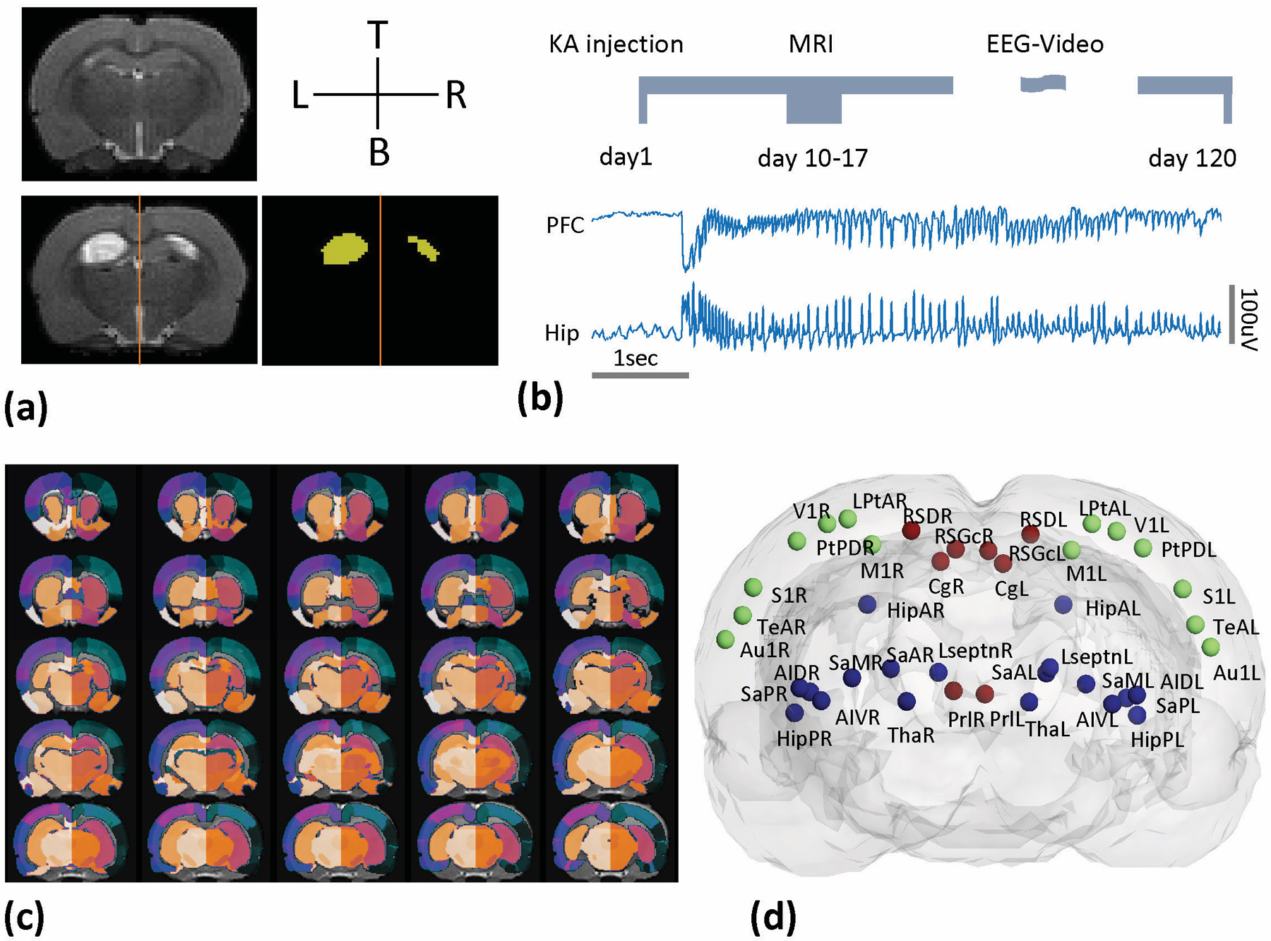
Eighteen rats with the intrahippocampal kainate model of mesial temporal lobe epilepsy were used for this experiment. Functional magnetic resonance imaging (fMRI) measurements were made 1 week after status epilepticus, followed by 2-4-month electrophysiological and video monitoring. Animals were identified as having (1) developed epilepsy (E+, n = 9) or (2) not developed epilepsy (E-, n = 6). Nine additional animals served as controls. Graph theory analysis was performed on the fMRI data to quantify the functional brain networks in all animals prior to the development of epilepsy. Spectrum clustering with the network features was performed to estimate their predictability in epileptogenesis.
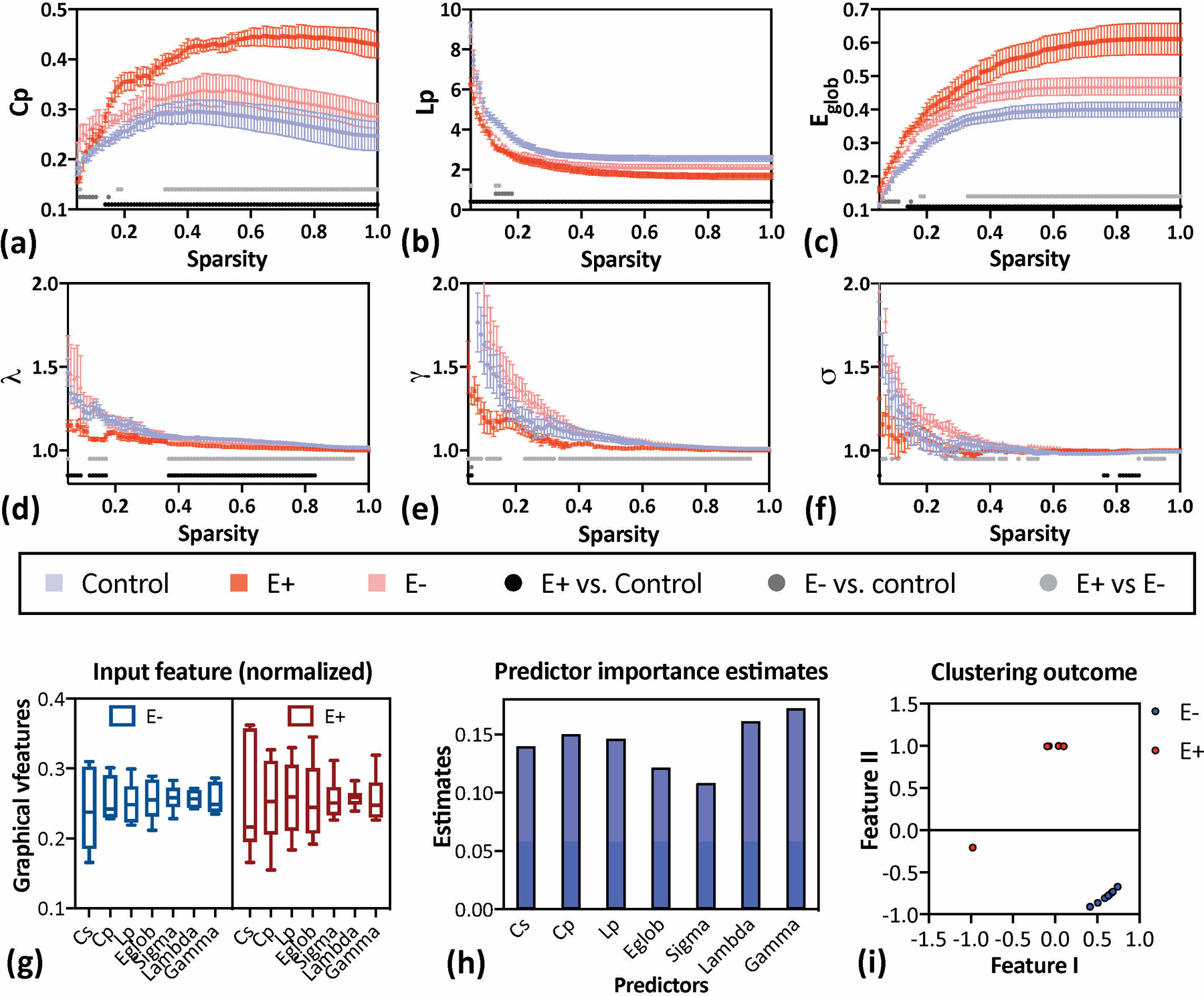
Our data indicated that E+ animals showed an overall increase in functional connectivity strength compared to E- and control animals. Global network features and small-worldness of E- rats were similar to controls, whereas E+ rats demonstrated increased small-worldness, including increased reorganization degree, clustering coefficient, and global efficiency, with reduced shortest pathlength. A notable classification of the combined brain network parameters was found in E+ and E- animals. For the local network parameters, the E- rats showed increased hubs in sensorimotor cortex, and decreased hubness in hippocampus. The E+ rats showed a complete loss of hippocampal hubs, and the appearance of new hubs in the prefrontal cortex. We also observed that lesion severity was not related to epileptogenesis.
Our data provide a view of the reorganization of topographical functional brain networks in the early period of epileptogenesis and how it can significantly predict the development of epilepsy. The differences from E- animals offer a potential means for applying noninvasive neuroimaging tools for the early prediction of epilepsy.
本研究旨在探讨癫痫发生早期的功能性脑网络表现。
本实验使用内侧颞叶癫痫海人酸模型的 18 只大鼠。在癫痫持续状态后 1 周进行功能磁共振成像(fMRI)测量,随后进行 2-4 个月的电生理和视频监测。动物被鉴定为(1)发展为癫痫(E+,n=9)或(2)未发展为癫痫(E-,n=6)。另外 9 只动物作为对照。在癫痫发生前,对所有动物的 fMRI 数据进行图论分析,以量化功能性脑网络。用网络特征进行谱聚类,以估计它们在癫痫发生中的可预测性。
我们的数据表明,E+动物的功能连接强度总体上高于 E-和对照组动物。E-大鼠的全局网络特征和小世界特性与对照组相似,而 E+大鼠表现出增加的小世界特性,包括增加的重组程度、聚类系数和全局效率,而最短路径长度降低。在 E+和 E-动物中发现了联合脑网络参数的显著分类。对于局部网络参数,E-大鼠在感觉运动皮层中显示出增加的枢纽,而在海马体中显示出减少的枢纽。E+大鼠表现出海马体枢纽完全丧失,而前额叶皮质出现新的枢纽。我们还观察到,损伤严重程度与癫痫发生无关。
我们的数据提供了癫痫发生早期拓扑功能脑网络重组的观点,以及它如何能够显著预测癫痫的发展。与 E-动物的差异为应用非侵入性神经影像学工具早期预测癫痫提供了一种潜在手段。