• 文献检索
  • 文档翻译
  • 深度研究
  • 学术资讯
  • Suppr Zotero 插件Zotero 插件
  • 邀请有礼
  • 套餐&价格
  • 历史记录
应用&插件
Suppr Zotero 插件Zotero 插件浏览器插件Mac 客户端Windows 客户端微信小程序
定价
高级版会员购买积分包购买API积分包
服务
文献检索文档翻译深度研究API 文档MCP 服务
关于我们
关于 Suppr公司介绍联系我们用户协议隐私条款
关注我们

Suppr 超能文献

核心技术专利:CN118964589B侵权必究
粤ICP备2023148730 号-1Suppr @ 2026

文献检索

告别复杂PubMed语法,用中文像聊天一样搜索,搜遍4000万医学文献。AI智能推荐,让科研检索更轻松。

立即免费搜索

文件翻译

保留排版,准确专业,支持PDF/Word/PPT等文件格式,支持 12+语言互译。

免费翻译文档

深度研究

AI帮你快速写综述,25分钟生成高质量综述,智能提取关键信息,辅助科研写作。

立即免费体验

将机器学习与纹理分析相结合,以区分肺癌患者的纵隔淋巴结。

Combining machine learning and texture analysis to differentiate mediastinal lymph nodes in lung cancer patients.

机构信息

Medical School, Sao Paulo State University Julio de Mesquita Filho, Botucatu, Brazil.

Institute of Bioscience, Sao Paulo State University Julio de Mesquita Filho, Botucatu, Brazil.

出版信息

Phys Eng Sci Med. 2021 Jun;44(2):387-394. doi: 10.1007/s13246-021-00988-2. Epub 2021 Mar 17.

DOI:10.1007/s13246-021-00988-2
PMID:33730292
原文链接:https://pmc.ncbi.nlm.nih.gov/articles/PMC7967117/
Abstract

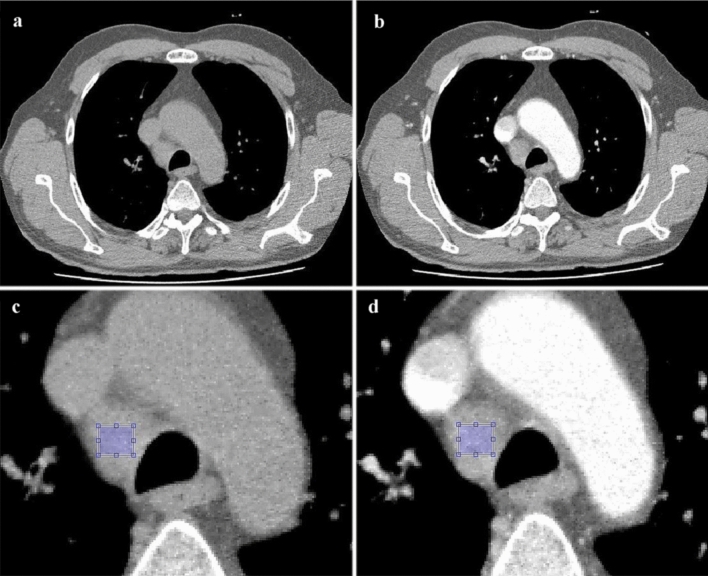
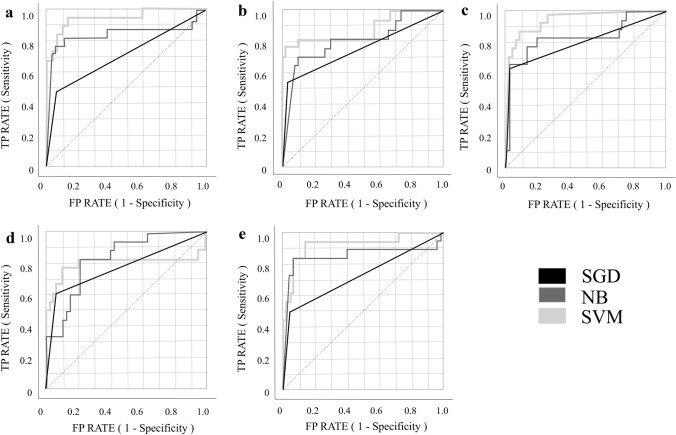
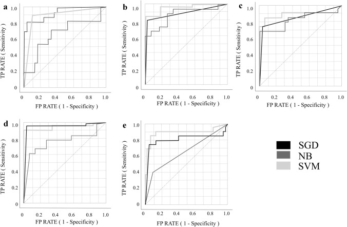
Evaluate whether texture analysis associated with machine learning approaches could differentiate between malignant and benign lymph nodes. A total 18 patients with lung cancer were selected, with 39 lymph nodes, being 15 malignant and 24 benign. Retrospective computed tomography scans were utilized both with and without contrast medium. The great differential of this work was the use of 15 textures from mediastinal lymph nodes, with five different physicians as operators. First and second order statistical textures such as gray level run length and co-occurrence matrix were extracted and applied to three different machine learning classifiers. The best machine learning classifier demonstrated a variability of less than 5% among operators. The support vector machine (SVM) classifier presented 95% of the area under the ROC curve (AUC) and 89% of sensitivity for sequences without contrast medium. SVM classifier presented 93% of AUC and 86% of sensitivity for sequences with contrast medium. Texture analysis and machine learning may be helpful in the differentiation between malign and benign lymph nodes. This study can aid the physician in diagnosis and staging of lymph nodes and potentially reduce the number of invasive analysis to histopathological confirmation.

摘要

评估纹理分析与机器学习方法相结合是否可以区分良恶性淋巴结。共选择了 18 例肺癌患者,共 39 个淋巴结,其中 15 个为恶性,24 个为良性。使用了有和没有对比剂的回顾性计算机断层扫描。这项工作的最大不同之处在于使用了来自纵隔淋巴结的 15 种纹理,有 5 位不同的医生作为操作者。提取了一阶和二阶统计纹理,如灰度游程长度和共生矩阵,并应用于三种不同的机器学习分类器。最佳的机器学习分类器在操作者之间的变异性小于 5%。支持向量机 (SVM) 分类器在无对比剂序列中呈现出 95%的ROC 曲线下面积(AUC)和 89%的敏感性。SVM 分类器在有对比剂的序列中呈现出 93%的 AUC 和 86%的敏感性。纹理分析和机器学习可能有助于区分良恶性淋巴结。这项研究可以帮助医生对淋巴结进行诊断和分期,并有可能减少对组织病理学确认的侵入性分析数量。

https://cdn.ncbi.nlm.nih.gov/pmc/blobs/e91e/7967117/2996bb0bdb3f/13246_2021_988_Fig3_HTML.jpg
https://cdn.ncbi.nlm.nih.gov/pmc/blobs/e91e/7967117/a5243a0f97ac/13246_2021_988_Fig1_HTML.jpg
https://cdn.ncbi.nlm.nih.gov/pmc/blobs/e91e/7967117/c8a86cd86692/13246_2021_988_Fig2_HTML.jpg
https://cdn.ncbi.nlm.nih.gov/pmc/blobs/e91e/7967117/2996bb0bdb3f/13246_2021_988_Fig3_HTML.jpg
https://cdn.ncbi.nlm.nih.gov/pmc/blobs/e91e/7967117/a5243a0f97ac/13246_2021_988_Fig1_HTML.jpg
https://cdn.ncbi.nlm.nih.gov/pmc/blobs/e91e/7967117/c8a86cd86692/13246_2021_988_Fig2_HTML.jpg
https://cdn.ncbi.nlm.nih.gov/pmc/blobs/e91e/7967117/2996bb0bdb3f/13246_2021_988_Fig3_HTML.jpg

相似文献

1
Combining machine learning and texture analysis to differentiate mediastinal lymph nodes in lung cancer patients.将机器学习与纹理分析相结合,以区分肺癌患者的纵隔淋巴结。
Phys Eng Sci Med. 2021 Jun;44(2):387-394. doi: 10.1007/s13246-021-00988-2. Epub 2021 Mar 17.
2
Quantitative CT texture and shape analysis: can it differentiate benign and malignant mediastinal lymph nodes in patients with primary lung cancer?定量CT纹理和形态分析:它能否区分原发性肺癌患者的良性和恶性纵隔淋巴结?
Eur Radiol. 2015 Feb;25(2):480-7. doi: 10.1007/s00330-014-3420-6. Epub 2014 Sep 13.
3
Texture Analysis and Synthesis of Malignant and Benign Mediastinal Lymph Nodes in Patients with Lung Cancer on Computed Tomography.肺癌患者 CT 图像中纵隔良恶性淋巴结的纹理分析与合成。
Sci Rep. 2017 Feb 24;7:43209. doi: 10.1038/srep43209.
4
Machine learning to identify lymph node metastasis from thyroid cancer in patients undergoing contrast-enhanced CT studies.机器学习在增强 CT 研究中识别甲状腺癌患者淋巴结转移的应用。
Radiography (Lond). 2021 Aug;27(3):920-926. doi: 10.1016/j.radi.2021.03.001. Epub 2021 Mar 21.
5
CT texture analysis can help differentiate between malignant and benign lymph nodes in the mediastinum in patients suspected for lung cancer.CT纹理分析有助于鉴别疑似肺癌患者纵隔内恶性和良性淋巴结。
Acta Radiol. 2016 Jun;57(6):669-76. doi: 10.1177/0284185115598808. Epub 2015 Aug 12.
6
The method and efficacy of support vector machine classifiers based on texture features and multi-resolution histogram from (18)F-FDG PET-CT images for the evaluation of mediastinal lymph nodes in patients with lung cancer.基于(18)F-FDG PET-CT图像纹理特征和多分辨率直方图的支持向量机分类器用于评估肺癌患者纵隔淋巴结的方法及疗效
Eur J Radiol. 2015 Feb;84(2):312-7. doi: 10.1016/j.ejrad.2014.11.006. Epub 2014 Nov 18.
7
Machine learning-based diagnostic method of pre-therapeutic F-FDG PET/CT for evaluating mediastinal lymph nodes in non-small cell lung cancer.基于机器学习的非小细胞肺癌治疗前 F-FDG PET/CT 纵隔淋巴结诊断方法。
Eur Radiol. 2021 Jun;31(6):4184-4194. doi: 10.1007/s00330-020-07523-z. Epub 2020 Nov 25.
8
Prediction of mediastinal lymph node metastasis based on F-FDG PET/CT imaging using support vector machine in non-small cell lung cancer.基于 F-FDG PET/CT 影像的支持向量机对非小细胞肺癌纵隔淋巴结转移的预测。
Eur Radiol. 2021 Jun;31(6):3983-3992. doi: 10.1007/s00330-020-07466-5. Epub 2020 Nov 17.
9
Radiomic Analysis using Density Threshold for FDG-PET/CT-Based N-Staging in Lung Cancer Patients.基于密度阈值的FDG-PET/CT对肺癌患者进行N分期的影像组学分析
Mol Imaging Biol. 2017 Apr;19(2):315-322. doi: 10.1007/s11307-016-0996-z.
10
Is SUVmax Helpful in the Differential Diagnosis of Enlarged Mediastinal Lymph Nodes? A Pilot Study.SUVmax 在纵隔淋巴结肿大的鉴别诊断中有帮助吗?一项初步研究。
Contrast Media Mol Imaging. 2018 Oct 28;2018:3417190. doi: 10.1155/2018/3417190. eCollection 2018.

引用本文的文献

1
Advancements and future trends in machine learning for lung cancer: a comprehensive bibliometric analysis.肺癌机器学习的进展与未来趋势:一项全面的文献计量分析
Clin Transl Oncol. 2025 Jun 4. doi: 10.1007/s12094-025-03945-7.
2
Enhanced lung cancer detection: Integrating improved random walker segmentation with artificial neural network and random forest classifier.增强型肺癌检测:将改进的随机游走分割与人工神经网络和随机森林分类器相结合。
Heliyon. 2024 Apr 4;10(7):e29032. doi: 10.1016/j.heliyon.2024.e29032. eCollection 2024 Apr 15.
3
Machine Learning and Texture Analysis of [F]FDG PET/CT Images for the Prediction of Distant Metastases in Non-Small-Cell Lung Cancer Patients.

本文引用的文献

1
Classification Models for Skin Tumor Detection Using Texture Analysis in Medical Images.基于医学图像纹理分析的皮肤肿瘤检测分类模型
J Imaging. 2020 Jun 19;6(6):51. doi: 10.3390/jimaging6060051.
2
CT-based radiomics for differentiating renal tumours: a systematic review.基于 CT 的放射组学在肾脏肿瘤鉴别中的应用:一项系统综述。
Abdom Radiol (NY). 2021 May;46(5):2052-2063. doi: 10.1007/s00261-020-02832-9. Epub 2020 Nov 2.
3
Covid-19: automatic detection from X-ray images utilizing transfer learning with convolutional neural networks.
用于预测非小细胞肺癌患者远处转移的[F]FDG PET/CT图像的机器学习与纹理分析
Biomedicines. 2024 Feb 20;12(3):472. doi: 10.3390/biomedicines12030472.
4
Radiomics analysis of bone marrow biopsy locations in [F]FDG PET/CT images for measurable residual disease assessment in multiple myeloma.基于[F]FDG PET/CT 图像骨髓活检部位的影像组学分析在多发性骨髓瘤中评估可测量残留疾病。
Phys Eng Sci Med. 2023 Jun;46(2):903-913. doi: 10.1007/s13246-023-01265-0. Epub 2023 May 8.
5
Investigation on the incidence and risk factors of lung cancer among Chinese hospital employees.中国医院职工肺癌发病率及危险因素调查。
Thorac Cancer. 2022 Aug;13(15):2210-2222. doi: 10.1111/1759-7714.14549. Epub 2022 Jul 11.
新冠病毒(Covid-19):利用卷积神经网络的迁移学习从 X 光图像中自动检测。
Phys Eng Sci Med. 2020 Jun;43(2):635-640. doi: 10.1007/s13246-020-00865-4. Epub 2020 Apr 3.
4
Machine learning analysis of MRI-derived texture features to predict placenta accreta spectrum in patients with placenta previa.基于 MRI 纹理特征的机器学习分析预测前置胎盘患者胎盘植入谱。
Magn Reson Imaging. 2019 Dec;64:71-76. doi: 10.1016/j.mri.2019.05.017. Epub 2019 May 15.
5
Texture Analysis of Imaging: What Radiologists Need to Know.医学影像学中的纹理分析:放射科医师须知。
AJR Am J Roentgenol. 2019 Mar;212(3):520-528. doi: 10.2214/AJR.18.20624. Epub 2019 Jan 15.
6
Prediction of quadriceps strength recovery after anterior cruciate ligament reconstruction with a hamstring autograft: Decision tree analysis.自体腘绳肌腱移植重建前交叉韧带后股四头肌力量恢复的预测:决策树分析
J Orthop Sci. 2019 Mar;24(2):301-305. doi: 10.1016/j.jos.2018.10.007. Epub 2018 Nov 2.
7
The 8 lung cancer TNM classification and clinical staging system: review of the changes and clinical implications.第八版肺癌TNM分类及临床分期系统:变化及临床意义综述
Quant Imaging Med Surg. 2018 Aug;8(7):709-718. doi: 10.21037/qims.2018.08.02.
8
Clear Cell Renal Cell Carcinoma: Machine Learning-Based Quantitative Computed Tomography Texture Analysis for Prediction of Fuhrman Nuclear Grade.透明细胞肾细胞癌:基于机器学习的定量 CT 纹理分析预测 Fuhrman 核分级。
Eur Radiol. 2019 Mar;29(3):1153-1163. doi: 10.1007/s00330-018-5698-2. Epub 2018 Aug 30.
9
Quantitative Contour Analysis as an Image-based Discriminator Between Benign and Malignant Renal Tumors.定量轮廓分析作为基于图像的肾良性与恶性肿瘤鉴别方法
Urology. 2018 Apr;114:121-127. doi: 10.1016/j.urology.2017.12.018. Epub 2018 Jan 2.
10
Benign and malignant enlarged chest nodes staging by diffusion-weighted MRI: an alternative to mediastinoscopy?通过扩散加权磁共振成像对良性和恶性胸部肿大淋巴结进行分期:纵隔镜检查的替代方法?
Br J Radiol. 2018 Feb;91(1082):20160919. doi: 10.1259/bjr.20160919. Epub 2017 Dec 18.