Sumi Koichiro, Otani Naoki, Mori Fumi, Yamamuro Shun, Oshima Hideki, Yoshino Atsuo
Division of Neurosurgery, Department of Neurological Surgery, Nihon University School of Medicine, 30-1 Oyaguchi-Kamimachi, Itabashi-ku, Tokyo, 173-8610, Japan.
BMC Neurol. 2021 Mar 17;21(1):119. doi: 10.1186/s12883-021-02144-5.
Intracranial venous hypertension has been associated with a few cases of meningioma secondary to compression of the venous sinus. This is the rare case of small meningioma involving the sigmoid sinus leading to intracranial venous hypertension mimicking venous thrombosis.
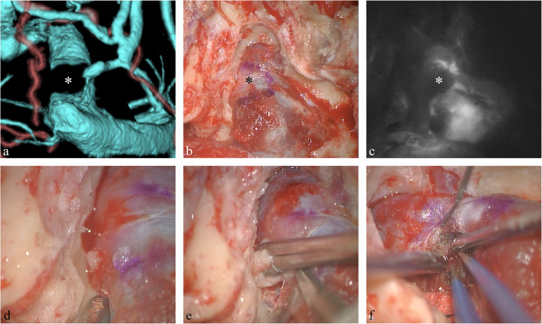
A 39-year-old woman suffered visual dysfunction due to bilateral papilledema. Noncontrast head computed tomography (CT) showed no intracranial space-occupying lesions or hydrocephalus. Cerebrospinal fluid examination revealed high opening pressure. Various image inspections such as three-dimensional CT angiography, magnetic resonance imaging, and cerebral angiography demonstrated a small 2.5-cm lesion causing subtotal occlusion of the dominant right sigmoid sinus. No improvement of clinical manifestations was observed after medical treatment for 6 months, so right presigmoid craniectomy was performed. Operative findings revealed that the tumor was located predominantly involving the sigmoid sinus. The pathological diagnosis was fibrous meningioma. Postoperative fundoscopic examination showed improvement of bilateral papilledema.
We treated a patient presenting with intracranial hypertension due to a small meningioma involving the sigmoid sinus. This unusual case suggests that early surgical strategies should be undertaken to relieve the sinus obstruction.
颅内静脉高压与少数因静脉窦受压继发的脑膜瘤病例有关。这是一例罕见的累及乙状窦的小型脑膜瘤导致颅内静脉高压并酷似静脉血栓形成的病例。
一名39岁女性因双侧视乳头水肿出现视觉功能障碍。头颅非增强计算机断层扫描(CT)未显示颅内占位性病变或脑积水。脑脊液检查显示初压升高。三维CT血管造影、磁共振成像和脑血管造影等各种影像学检查显示一个2.5厘米的小病变导致右侧优势乙状窦几乎完全闭塞。药物治疗6个月后临床表现无改善,因此进行了右侧乙状窦前颅骨切除术。手术发现肿瘤主要位于乙状窦。病理诊断为纤维性脑膜瘤。术后眼底检查显示双侧视乳头水肿有所改善。
我们治疗了一名因累及乙状窦的小型脑膜瘤而出现颅内高压的患者。这个不寻常的病例表明应尽早采取手术策略以解除窦道阻塞。