• 文献检索
  • 文档翻译
  • 深度研究
  • 学术资讯
  • Suppr Zotero 插件Zotero 插件
  • 邀请有礼
  • 套餐&价格
  • 历史记录
应用&插件
Suppr Zotero 插件Zotero 插件浏览器插件Mac 客户端Windows 客户端微信小程序
定价
高级版会员购买积分包购买API积分包
服务
文献检索文档翻译深度研究API 文档MCP 服务
关于我们
关于 Suppr公司介绍联系我们用户协议隐私条款
关注我们

Suppr 超能文献

核心技术专利:CN118964589B侵权必究
粤ICP备2023148730 号-1Suppr @ 2026

文献检索

告别复杂PubMed语法,用中文像聊天一样搜索,搜遍4000万医学文献。AI智能推荐,让科研检索更轻松。

立即免费搜索

文件翻译

保留排版,准确专业,支持PDF/Word/PPT等文件格式,支持 12+语言互译。

免费翻译文档

深度研究

AI帮你快速写综述,25分钟生成高质量综述,智能提取关键信息,辅助科研写作。

立即免费体验

踝关节管内胫神经及其分支的 MRI 研究:一种应用 3D-FIESTA-C 序列进行多平面重建的方法。

An MRI study of the tibial nerve in the ankle canal and its branches: a method of multiplanar reformation with 3D-FIESTA-C sequences.

机构信息

Department of Radiology, Shandong Mental Health Center, Shandong, 250014, China.

Department of Radiology, Third Medical Centre of Chinese PLA General Hospital, Beijing, 100039, China.

出版信息

BMC Med Imaging. 2021 Mar 17;21(1):51. doi: 10.1186/s12880-021-00582-8.

DOI:10.1186/s12880-021-00582-8
PMID:33731040
原文链接:https://pmc.ncbi.nlm.nih.gov/articles/PMC7968234/
Abstract

BACKGROUND

The visualization of the tibial nerve and its branches in the ankle canal is helpful for the diagnosis of local lesions and compression, and it is also useful for clinical observation and surgical planning. The aim of this study was to investigate the feasibility of three-dimensional dual-excitation balanced steady-state free precession sequence (3D-FIESTA-C) multiplanar reformation (MPR) display of the tibial nerve and its branches in the ankle canal.

METHODS

The subjects were 20 healthy volunteers (40 ankles), aged 22-50 years, with no history of ankle joint disease. The 3D-FIESTA-C sequence was used in the 3.0 T magnetic resonance equipment for imaging. During scanning, each foot was at an angle of 90° to the tibia. The tibial nerve of the ankle canal and its branches were displayed and measured at the same level through MPR.

RESULTS

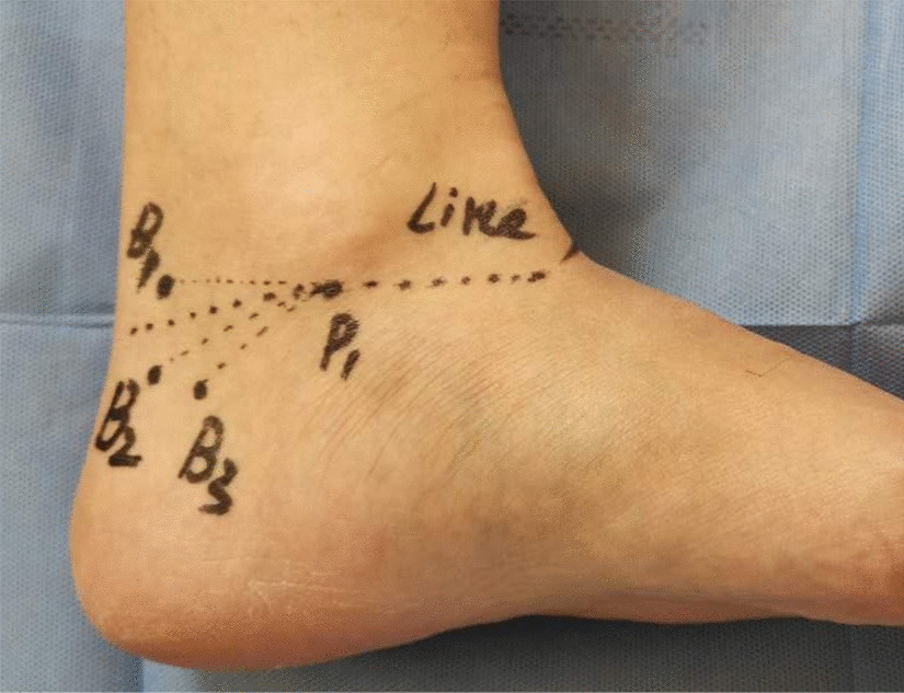
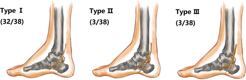
Most of the tibial nerve bifurcation points were located in the ankle canal (57.5%), few bifurcation points (42.5%) were located at the proximal end of the ankle canal, and none of them were found away from the distal end. The bifurcation between the medial plantar nerve and the lateral plantar nerve was on the line between the tip of the medial malleolus and the calcaneus, and it's angle ranged between 6° and 35°. In MPR images, the display rates of both the medial calcaneal nerve and the subcalcaneal nerve were 100%, and the starting point of the subcalcaneal nerve was always at the distal end of the starting point of the medial calcaneal nerve. In 55% of cases, there were more than two medial calcaneal nerve innervations.

CONCLUSION

The 3D-FIESTA-C MPR can display the morphological features and positions of the tibial nerve and its branches and the bifurcation point's projection position can be marked on the body surface. This method not only benefited the imaging diagnosis of the tibial nerve and branch-related lesions in the ankle canal, but it also provided a good imaging basis to plan a clinical operation of the ankle canal and avoid surgical injury.

摘要

背景

在踝关节管内可视化胫神经及其分支有助于诊断局部病变和压迫,也有助于临床观察和手术规划。本研究旨在探讨三维双激发平衡稳态自由进动序列(3D-FIESTA-C)多平面重建(MPR)显示踝关节管内胫神经及其分支的可行性。

方法

本研究纳入 20 名健康志愿者(40 侧踝关节),年龄 22-50 岁,均无踝关节疾病史。使用 3.0T 磁共振设备进行 3D-FIESTA-C 序列成像。扫描时,每只脚与胫骨成 90°角。在 MPR 上同一水平显示和测量踝关节管内胫神经及其分支。

结果

大多数胫神经分叉点位于踝关节管内(57.5%),少数分叉点(42.5%)位于踝关节管近端,无分叉点位于踝关节管远端。内侧足底神经和外侧足底神经的分叉点位于内踝尖和跟骨之间的连线上,角度在 6°-35°之间。MPR 图像中,内侧跟骨神经和跗骨下神经的显示率均为 100%,跗骨下神经的起点始终位于内侧跟骨神经起点的远端。在 55%的情况下,有超过两条内侧跟骨神经支配。

结论

3D-FIESTA-C MPR 可显示胫神经及其分支的形态特征和位置,可在体表标记分叉点的投影位置。该方法不仅有助于诊断踝关节管内胫神经及其分支相关病变的影像学表现,而且为踝关节管的临床手术规划提供了良好的影像学依据,避免了手术损伤。

https://cdn.ncbi.nlm.nih.gov/pmc/blobs/7f9a/7968234/587aa2f7458c/12880_2021_582_Fig12_HTML.jpg
https://cdn.ncbi.nlm.nih.gov/pmc/blobs/7f9a/7968234/7b5165abae36/12880_2021_582_Fig1_HTML.jpg
https://cdn.ncbi.nlm.nih.gov/pmc/blobs/7f9a/7968234/564f67ea756f/12880_2021_582_Fig2_HTML.jpg
https://cdn.ncbi.nlm.nih.gov/pmc/blobs/7f9a/7968234/2d8bdb6d0750/12880_2021_582_Fig3_HTML.jpg
https://cdn.ncbi.nlm.nih.gov/pmc/blobs/7f9a/7968234/8f99019a2489/12880_2021_582_Fig4_HTML.jpg
https://cdn.ncbi.nlm.nih.gov/pmc/blobs/7f9a/7968234/8b2757c635ec/12880_2021_582_Fig5_HTML.jpg
https://cdn.ncbi.nlm.nih.gov/pmc/blobs/7f9a/7968234/3a774de1082d/12880_2021_582_Fig6_HTML.jpg
https://cdn.ncbi.nlm.nih.gov/pmc/blobs/7f9a/7968234/214946a21d51/12880_2021_582_Fig7_HTML.jpg
https://cdn.ncbi.nlm.nih.gov/pmc/blobs/7f9a/7968234/5a0165d82495/12880_2021_582_Fig8_HTML.jpg
https://cdn.ncbi.nlm.nih.gov/pmc/blobs/7f9a/7968234/9ccc1be1830a/12880_2021_582_Fig9_HTML.jpg
https://cdn.ncbi.nlm.nih.gov/pmc/blobs/7f9a/7968234/bafbdb99f1cb/12880_2021_582_Fig10_HTML.jpg
https://cdn.ncbi.nlm.nih.gov/pmc/blobs/7f9a/7968234/686d91c4b379/12880_2021_582_Fig11_HTML.jpg
https://cdn.ncbi.nlm.nih.gov/pmc/blobs/7f9a/7968234/587aa2f7458c/12880_2021_582_Fig12_HTML.jpg

相似文献

1
An MRI study of the tibial nerve in the ankle canal and its branches: a method of multiplanar reformation with 3D-FIESTA-C sequences.踝关节管内胫神经及其分支的 MRI 研究:一种应用 3D-FIESTA-C 序列进行多平面重建的方法。
BMC Med Imaging. 2021 Mar 17;21(1):51. doi: 10.1186/s12880-021-00582-8.
2
Ultrasound-guided topographic anatomy of the medial calcaneal branches of the tibial nerve.超声引导下胫神经内踝后支的局部解剖学。
Folia Morphol (Warsz). 2021;80(2):267-274. doi: 10.5603/FM.a2020.0062. Epub 2020 Jun 3.
3
Morphometric assessment of tibial nerve and its branches around the ankle.踝周胫神经及其分支的形态计量评估。
Medicine (Baltimore). 2024 Apr 12;103(15):e37745. doi: 10.1097/MD.0000000000037745.
4
The rotating stretched curved planar reconstruction of 3D-FIESTA MR imaging for evaluating the anterior cruciate ligament of the knee joint.用于评估膝关节前交叉韧带的3D-FIESTA磁共振成像的旋转拉伸曲面重建
Magn Reson Imaging. 2019 Jan;55:46-51. doi: 10.1016/j.mri.2018.09.013. Epub 2018 Sep 14.
5
Normal Anatomy and Compression Areas of Nerves of the Foot and Ankle: US and MR Imaging with Anatomic Correlation.足踝部神经的正常解剖及受压区域:超声与磁共振成像及解剖学对照
Radiographics. 2015 Sep-Oct;35(5):1469-82. doi: 10.1148/rg.2015150028. Epub 2015 Aug 18.
6
3D-FIESTA MRI at 3 T demonstrating branches of the intraparotid facial nerve, parotid ducts and relation with benign parotid tumours.3T 下的 3D-FIESTA MRI 显示腮腺内面神经分支、腮腺导管及其与良性腮腺肿瘤的关系。
Clin Radiol. 2012 Nov;67(11):1078-82. doi: 10.1016/j.crad.2012.03.014. Epub 2012 May 16.
7
Variations in the branching pattern of tibial nerve in foot: a review of literature and relevant clinical anatomy.足部胫神经分支模式的变化:文献复习与相关临床解剖学
Folia Morphol (Warsz). 2023;82(2):231-241. doi: 10.5603/FM.a2022.0042. Epub 2022 Apr 28.
8
Morphological evaluation of the calcaneofibular ligament in different ankle positions using a three-dimensional MRI sequence.使用三维MRI序列对不同踝关节位置的跟腓韧带进行形态学评估。
Surg Radiol Anat. 2019 Mar;41(3):307-311. doi: 10.1007/s00276-018-2152-8. Epub 2018 Dec 11.
9
The medial and inferior calcaneal nerves: an anatomic study.跟骨内侧和下神经:一项解剖学研究。
Surg Radiol Anat. 1999;21(3):169-73. doi: 10.1007/BF01630895.
10
The posteromedial neurovascular bundle of the ankle: an anatomic study using plastinated cross sections.踝关节的后内侧神经血管束:一项使用塑化横断面的解剖学研究
Arthroscopy. 2008 Mar;24(3):258-263.e1. doi: 10.1016/j.arthro.2007.08.030. Epub 2007 Nov 9.

引用本文的文献

1
An Anatomical Study on the Tibial Nerve Bifurcation Level in Relation to the Malleolar-Calcaneal Axis and Its Clinical Significance.胫神经分叉水平与踝-跟骨轴线关系的解剖学研究及其临床意义
Cureus. 2025 Jun 21;17(6):e86511. doi: 10.7759/cureus.86511. eCollection 2025 Jun.
2
Navigating the anomalous path of the tibial nerve at the ankle - attention to the intricacies!探寻踝关节处胫神经的异常走行——关注其中的错综复杂之处!
Indian J Anaesth. 2024 Mar;68(3):307-308. doi: 10.4103/ija.ija_1126_23. Epub 2024 Feb 22.
3
The role of high-resolution ultrasound and MRI in the evaluation of peripheral nerves in the lower extremity.

本文引用的文献

1
The injection site in the tarsal tunnel to minimize neurovascular injury for heel pain: an anatomical study.为了减轻足跟痛神经血管损伤,在跗管内注射:一项解剖学研究。
Surg Radiol Anat. 2020 Jun;42(6):681-684. doi: 10.1007/s00276-019-02411-8. Epub 2020 Jan 14.
2
Technological Advancements in Magnetic Resonance Neurography.磁共振神经成像技术的进展。
Curr Neurol Neurosci Rep. 2019 Aug 24;19(10):75. doi: 10.1007/s11910-019-0996-x.
3
Interfascicular septum of the calcaneal tunnel and its relationship with the plantar nerves: A cadaveric study.
高分辨率超声和磁共振成像在评估下肢周围神经中的作用。
J Ultrason. 2023 Nov 23;23(95):e328-e346. doi: 10.15557/jou.2023.0038. eCollection 2023 Oct.
4
3D isotropic MRI of ankle: review of literature with comparison to 2D MRI.踝关节 3D 各向同性 MRI:与 2D MRI 的文献综述比较。
Skeletal Radiol. 2024 May;53(5):825-846. doi: 10.1007/s00256-023-04513-2. Epub 2023 Nov 18.
5
Anatomical variations of the tibial nerve and their clinical correlation.胫神经的解剖变异及其临床关联。
Anat Cell Biol. 2023 Dec 31;56(4):415-420. doi: 10.5115/acb.23.065. Epub 2023 Sep 11.
跟腱管间隔及其与足底神经的关系:尸体研究。
Clin Anat. 2019 Oct;32(7):877-882. doi: 10.1002/ca.23381. Epub 2019 Apr 15.
4
[Clinical analysis of ankle arthroscopy technique for treatment of tarsal tunnel syndrome].踝关节镜技术治疗跗管综合征的临床分析
Zhonghua Yi Xue Za Zhi. 2018 Oct 9;98(37):2995-2998. doi: 10.3760/cma.j.issn.0376-2491.2018.37.009.
5
Clinical-anatomic mapping of the tarsal tunnel with regard to Baxter's neuropathy in recalcitrant heel pain syndrome: part I.顽固性足跟痛综合征中与巴克斯特神经病变相关的跗管临床解剖图谱:第一部分
Surg Radiol Anat. 2019 Jan;41(1):29-41. doi: 10.1007/s00276-018-2124-z. Epub 2018 Oct 27.
6
MRI of Foot Drop: How We Do It.足部下垂的 MRI:我们如何进行。
Radiology. 2018 Oct;289(1):9-24. doi: 10.1148/radiol.2018172634. Epub 2018 Aug 28.
7
Trifurcation of the tibial nerve within the tarsal tunnel.胫神经在跗管内的三叉分支。
Surg Radiol Anat. 2018 May;40(5):529-532. doi: 10.1007/s00276-017-1948-2. Epub 2017 Nov 24.
8
Peripheral nerve diffusion tensor imaging: Overview, pitfalls, and future directions.周围神经弥散张量成像:概述、陷阱和未来方向。
J Magn Reson Imaging. 2018 May;47(5):1171-1189. doi: 10.1002/jmri.25876. Epub 2017 Oct 30.
9
Rate of Neurologic Injury Following Lateralizing Calcaneal Osteotomy Performed Through a Medial Approach.经内侧入路行跟骨外侧截骨术后的神经损伤发生率
Foot Ankle Int. 2017 Dec;38(12):1367-1373. doi: 10.1177/1071100717728678. Epub 2017 Sep 1.
10
Medial calcaneal neuropathy: A rare cause of prolonged heel pain.跟骨内侧神经病变:足跟长期疼痛的罕见原因。
Agri. 2017 Jan;29(1):43-46. doi: 10.5505/agri.2015.13540.