Department of Diagnostic and Interventional Radiology, Division of Cardiothoracic and Vascular Imaging, Lausanne University Hospital (CHUV), Rue du Bugnon 46, 1011, Lausanne, Switzerland.
Faculty of Biology and Medicine (FBM), University of Lausanne (UNIL), Lausanne, Switzerland.
Eur Radiol. 2021 Sep;31(9):7132-7142. doi: 10.1007/s00330-021-07809-w. Epub 2021 Mar 19.
To quantitatively evaluate the impact of virtual monochromatic images (VMI) on reduced-iodine-dose dual-energy coronary computed tomography angiography (CCTA) in terms of coronary lumen segmentation in vitro, and secondly to assess the image quality in vivo, compared with conventional CT obtained with regular iodine dose.
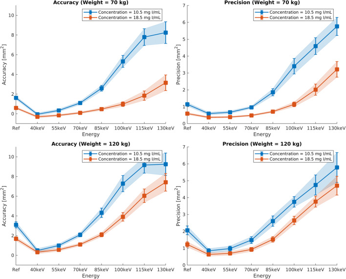
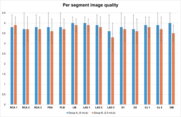
A phantom simulating regular and reduced iodine injection was used to determine the accuracy and precision of lumen area segmentation for various VMI energy levels. We retrospectively included 203 patients from December 2017 to August 2018 (mean age, 51.7 ± 16.8 years) who underwent CCTA using either standard (group A, n = 103) or reduced (group B, n = 100) iodine doses. Conventional images (group A) were qualitatively and quantitatively compared with 55-keV VMI (group B). We recorded the location of venous catheters.
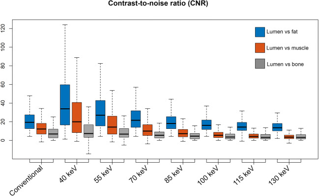
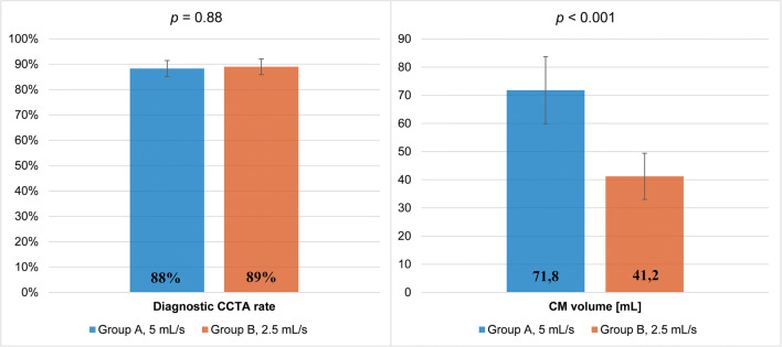
In vitro, VMI outperformed conventional CT, with a segmentation accuracy of 0.998 vs. 1.684 mm, respectively (p < 0.001), and a precision of 0.982 vs. 1.229 mm, respectively (p < 0.001), in simulated overweight adult subjects. In vivo, the rate of diagnostic CCTA in groups A and B was 88.4% (n = 91/103) vs. 89% (n = 89/100), respectively, and noninferiority of protocol B was inferred. Contrast-to-noise ratios (CNR) of lumen versus fat and muscle were higher in group B (p < 0.001) and comparable for lumen versus calcium (p = 0.423). Venous catheters were more often placed on the forearm or hand in group B (p < 0.001).
In vitro, low-keV VMI improve vessel area segmentation. In vivo, low-keV VMI allows for a 40% iodine dose and injection rate reduction while maintaining diagnostic image quality and improves the CNR between lumen versus fat and muscle.
• Dual-energy coronary CT angiography is becoming increasingly available and might help improve patient management. • Compared with regular-iodine-dose coronary CT angiography, reduced-iodine-dose dual-energy CT with low-keV monochromatic image reconstructions performed better in phantom-based vessel cross-sectional segmentation and proved to be noninferior in vivo. • Patients receiving reduced-iodine-dose dual-energy coronary CT angiography often had the venous catheter placed on the forearm or wrist without compromising image quality.
通过离体冠状动脉管腔分割评估虚拟单能量图像(VMI)对减少碘剂量双能冠状动脉 CT 血管造影(CCTA)的影响,并与常规碘剂量获得的常规 CT 进行比较,评估体内图像质量。
使用模拟常规和减少碘注射的体模来确定各种 VMI 能量水平下管腔面积分割的准确性和精度。我们回顾性地纳入了 2017 年 12 月至 2018 年 8 月期间接受 CCTA 检查的 203 例患者(平均年龄 51.7±16.8 岁),这些患者分别接受了标准碘剂量(A 组,n=103)或减少碘剂量(B 组,n=100)。对常规图像(A 组)与 55keV VMI(B 组)进行定性和定量比较。我们记录了静脉导管的位置。
在体外,VMI 优于常规 CT,在模拟超重成年患者中,分割准确性分别为 0.998 对 1.684mm(p<0.001),精度分别为 0.982 对 1.229mm(p<0.001)。在体内,A 组和 B 组的诊断性 CCTA 率分别为 88.4%(n=91/103)和 89%(n=89/100),推断出 B 组方案的非劣效性。管腔与脂肪和肌肉的对比噪声比(CNR)在 B 组中更高(p<0.001),而管腔与钙的 CNR 相当(p=0.423)。B 组中静脉导管更常放置在前臂或手上(p<0.001)。
在体外,低 keV VMI 可改善血管面积分割。在体内,低 keV VMI 可使碘剂量和注射率降低 40%,同时保持诊断图像质量,并提高管腔与脂肪和肌肉之间的 CNR。
• 双能冠状动脉 CT 血管造影术越来越普及,可能有助于改善患者管理。• 与常规碘剂量冠状动脉 CT 血管造影相比,低 keV 单能量图像重建的低碘剂量双能 CT 在基于体模的血管横断面分割中表现更好,并且在体内证明是等效的。• 接受低碘剂量双能冠状动脉 CT 血管造影的患者通常将静脉导管放置在前臂或手腕上,而不会影响图像质量。