• 文献检索
  • 文档翻译
  • 深度研究
  • 学术资讯
  • Suppr Zotero 插件Zotero 插件
  • 邀请有礼
  • 套餐&价格
  • 历史记录
应用&插件
Suppr Zotero 插件Zotero 插件浏览器插件Mac 客户端Windows 客户端微信小程序
定价
高级版会员购买积分包购买API积分包
服务
文献检索文档翻译深度研究API 文档MCP 服务
关于我们
关于 Suppr公司介绍联系我们用户协议隐私条款
关注我们

Suppr 超能文献

核心技术专利:CN118964589B侵权必究
粤ICP备2023148730 号-1Suppr @ 2026

文献检索

告别复杂PubMed语法,用中文像聊天一样搜索,搜遍4000万医学文献。AI智能推荐,让科研检索更轻松。

立即免费搜索

文件翻译

保留排版,准确专业,支持PDF/Word/PPT等文件格式,支持 12+语言互译。

免费翻译文档

深度研究

AI帮你快速写综述,25分钟生成高质量综述,智能提取关键信息,辅助科研写作。

立即免费体验

弥散张量成像显示了脑白质高信号的不同病理过程。

Diffusion tensor imaging revealed different pathological processes of white matter hyperintensities.

机构信息

Department of Radiology, The Affiliated Yixing Hospital of Jiangsu University, NO.75 Tongzhenguan Road, Yixing, Jiangsu Province, 214200, P.R. China.

Department of Radiology, First Affiliated Hospital of Xi'an Jiaotong University, Xi'an, Shaanxi Province, China.

出版信息

BMC Neurol. 2021 Mar 19;21(1):128. doi: 10.1186/s12883-021-02140-9.

DOI:10.1186/s12883-021-02140-9
PMID:33740898
原文链接:https://pmc.ncbi.nlm.nih.gov/articles/PMC7977583/
Abstract

BACKGROUND

Although increasing evidence showed the correlations between white matter hyperintensities (WMHs) and cognitive impairment, the relationship between them is still modest. Many researchers began to focus on the variation caused by the heterogeneity of WMH. We tried to explore the pathological heterogeneity in WMH by using diffusion tensor imaging (DTI), so as to provide a new insight into the future research.

METHODS

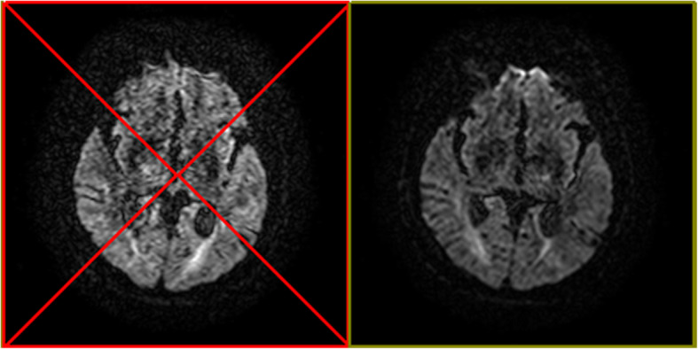
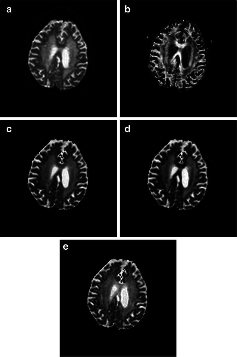
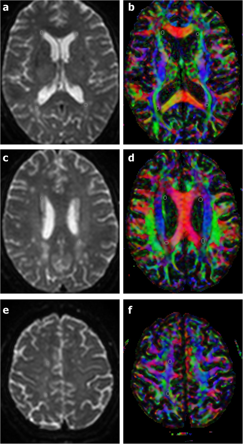
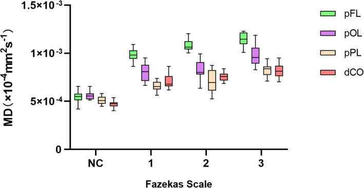
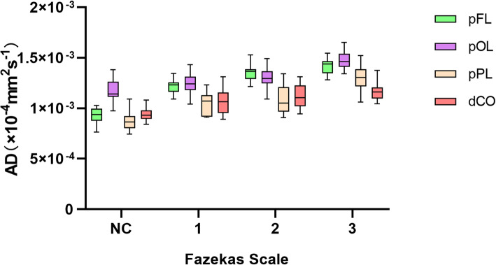
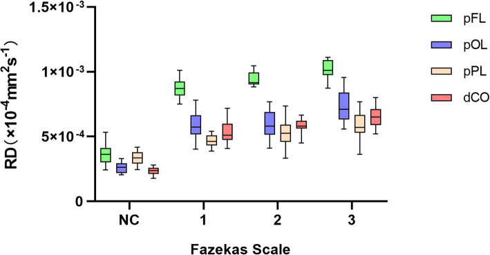
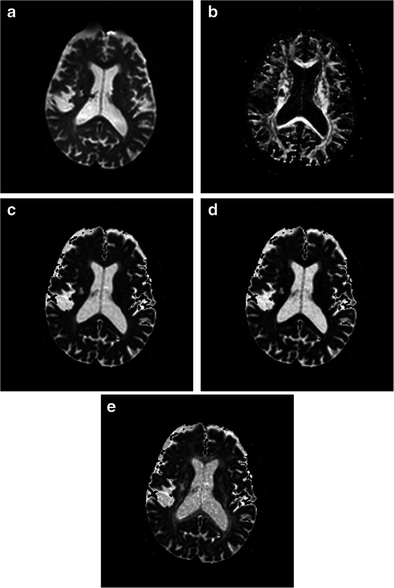
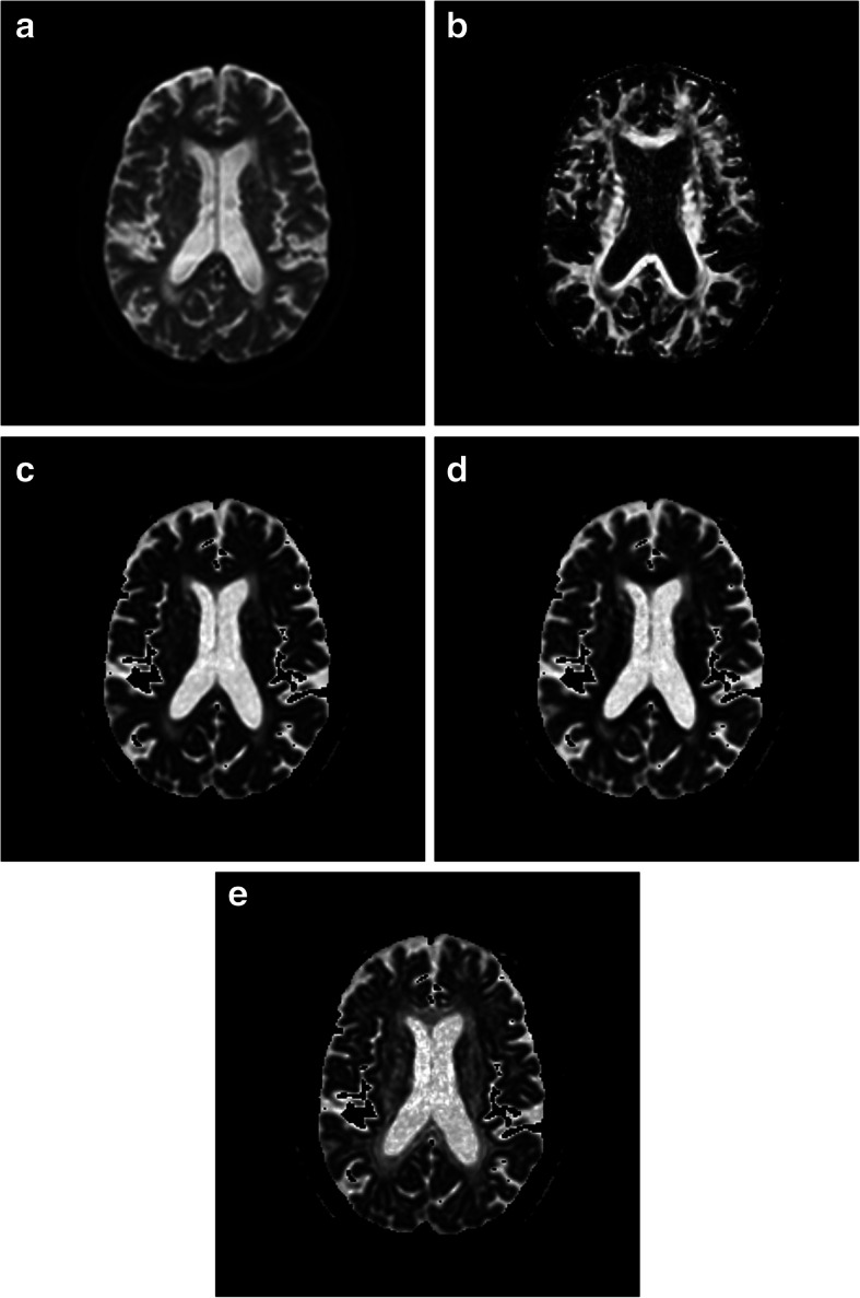
Diffusion weighted images (DWIs) of the brain were acquired from 73 patients with WMH and 18 healthy controls, which were then modeled by DTI. We measured fractional anisotropy (FA), mean diffusivity (MD), axial diffusivity (AD), and radial diffusivity (RD) of white matter of the periventricular frontal lobe (pFL), periventricular occipital lobe (pOL), periventricular parietal lobe (pPL) and deep centrum ovales (dCO), and grouped these measures according to the Fazekas scale. Then we compared the DTI metrics of different regions with the same Fazekas scale grade.

RESULTS

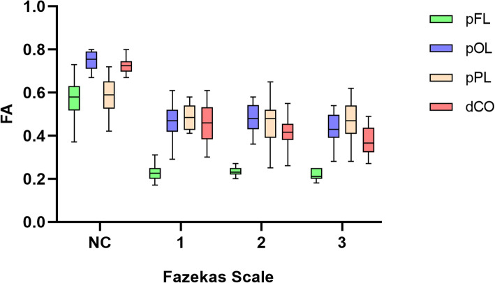
Significantly lower FA values (all p < 0.001), and higher MD (all p < 0.001) and RD values (all p < 0.001) were associated with WMH observed in the periventricular frontal lobe (pFL) compared to all other regions with the same Fazekas grades. The AD of WMH in the pFL was higher than that of pPL and dCO, but the differences between groups was not as high as of MD and RD, as indicated by the effect size. In the normal control group, DTI metrics between pFL and other regions were not significantly different or less significant different. The difference of DTI metrics of WMH between pPL, pOL and dCO was lower than that of normal white matter, as indicated by the effect size.

CONCLUSION

Distinct pathological processes can be revealed by DTI between frontal periventricular WMH and other regions. These processes may represent the effects of severe demyelination within the frontal periventricular WMH.

摘要

背景

尽管越来越多的证据表明脑白质高信号(WMH)与认知障碍之间存在相关性,但它们之间的关系仍然不太明确。许多研究人员开始关注WMH 异质性引起的变化。我们试图通过弥散张量成像(DTI)来探索 WMH 的病理异质性,以期为未来的研究提供新的视角。

方法

对 73 例 WMH 患者和 18 例健康对照者的脑部弥散加权图像(DWIs)进行采集,并通过 DTI 对其进行建模。我们测量了脑室周围额叶(pFL)、脑室周围枕叶(pOL)、脑室周围顶叶(pPL)和深部脑白质(dCO)的白质各向异性分数(FA)、平均弥散度(MD)、轴向弥散度(AD)和径向弥散度(RD),并根据 Fazekas 量表对这些指标进行分组。然后,我们比较了不同区域与相同 Fazekas 分级的 DTI 指标。

结果

与其他具有相同 Fazekas 分级的区域相比,脑室周围额叶(pFL)WMH 观察到的 FA 值显著降低(均 p<0.001),MD 值(均 p<0.001)和 RD 值(均 p<0.001)更高。与 pPL 和 dCO 相比,pFL 中 WMH 的 AD 值更高,但组间差异不如 MD 和 RD 显著,如效应大小所示。在正常对照组中,pFL 与其他区域之间的 DTI 指标没有显著差异或差异较小。与正常白质相比,pPL、pOL 和 dCO 之间 WMH 的 DTI 指标差异较小,如效应大小所示。

结论

DTI 可以揭示脑室周围额状 WM 与其他区域之间的不同病理过程。这些过程可能代表了脑室周围额状 WM 内严重脱髓鞘的影响。

https://cdn.ncbi.nlm.nih.gov/pmc/blobs/cc81/7977583/b88ba89b948d/12883_2021_2140_Fig9_HTML.jpg
https://cdn.ncbi.nlm.nih.gov/pmc/blobs/cc81/7977583/d44c2ee3c916/12883_2021_2140_Fig1_HTML.jpg
https://cdn.ncbi.nlm.nih.gov/pmc/blobs/cc81/7977583/94c20ff386ec/12883_2021_2140_Fig2_HTML.jpg
https://cdn.ncbi.nlm.nih.gov/pmc/blobs/cc81/7977583/4e0cf165f69b/12883_2021_2140_Fig3_HTML.jpg
https://cdn.ncbi.nlm.nih.gov/pmc/blobs/cc81/7977583/908f28c5ca7c/12883_2021_2140_Fig4_HTML.jpg
https://cdn.ncbi.nlm.nih.gov/pmc/blobs/cc81/7977583/2b0dc02dfb0e/12883_2021_2140_Fig5_HTML.jpg
https://cdn.ncbi.nlm.nih.gov/pmc/blobs/cc81/7977583/76394ad63851/12883_2021_2140_Fig6_HTML.jpg
https://cdn.ncbi.nlm.nih.gov/pmc/blobs/cc81/7977583/3c76e0ec2038/12883_2021_2140_Fig7_HTML.jpg
https://cdn.ncbi.nlm.nih.gov/pmc/blobs/cc81/7977583/2b5f57ea50b6/12883_2021_2140_Fig8_HTML.jpg
https://cdn.ncbi.nlm.nih.gov/pmc/blobs/cc81/7977583/b88ba89b948d/12883_2021_2140_Fig9_HTML.jpg

相似文献

1
Diffusion tensor imaging revealed different pathological processes of white matter hyperintensities.弥散张量成像显示了脑白质高信号的不同病理过程。
BMC Neurol. 2021 Mar 19;21(1):128. doi: 10.1186/s12883-021-02140-9.
2
Hemispheric Regional Based Analysis of Diffusion Tensor Imaging and Diffusion Tensor Tractography in Patients with Temporal Lobe Epilepsy and Correlation with Patient outcomes.基于半球区域的分析弥散张量成像和弥散张量纤维束成像在颞叶癫痫患者中的应用及其与患者预后的相关性。
Sci Rep. 2019 Jan 18;9(1):215. doi: 10.1038/s41598-018-36818-x.
3
A multimodal MRI approach to identify and characterize microstructural brain changes in neuropsychiatric systemic lupus erythematosus.一种用于识别和表征神经精神性系统性红斑狼疮患者脑微结构变化的多模态磁共振成像方法。
Neuroimage Clin. 2015 May 16;8:337-44. doi: 10.1016/j.nicl.2015.05.002. eCollection 2015.
4
Progression of White Matter Hyperintensities Preceded by Heterogeneous Decline of Microstructural Integrity.白质高信号进展之前存在微观结构完整性的异质性下降。
Stroke. 2018 Jun;49(6):1386-1393. doi: 10.1161/STROKEAHA.118.020980. Epub 2018 May 3.
5
Differences in Gaussian diffusion tensor imaging and non-Gaussian diffusion kurtosis imaging model-based estimates of diffusion tensor invariants in the human brain.基于高斯扩散张量成像和非高斯扩散峰度成像模型的人脑扩散张量不变量估计差异。
Med Phys. 2016 May;43(5):2464. doi: 10.1118/1.4946819.
6
Performances of diffusion kurtosis imaging and diffusion tensor imaging in detecting white matter abnormality in schizophrenia.扩散峰度成像和扩散张量成像在检测精神分裂症白质异常中的表现。
Neuroimage Clin. 2014 Dec 9;7:170-6. doi: 10.1016/j.nicl.2014.12.008. eCollection 2015.
7
Diffusion tensor imaging study of pediatric patients with congenital hydrocephalus: 1-year postsurgical outcomes.先天性脑积水患儿的扩散张量成像研究:术后1年的结果
J Neurosurg Pediatr. 2016 Sep;18(3):306-19. doi: 10.3171/2016.2.PEDS15628. Epub 2016 May 20.
8
White matter alterations and their associations with motor function in young adults born preterm with very low birth weight.早产儿极低出生体重儿成年后白质改变及其与运动功能的关系。
Neuroimage Clin. 2017 Oct 4;17:241-250. doi: 10.1016/j.nicl.2017.10.006. eCollection 2018.
9
Comparing a diffusion tensor and non-tensor approach to white matter fiber tractography in chronic stroke.比较扩散张量成像和非张量成像方法在慢性卒中白质纤维束成像中的应用
Neuroimage Clin. 2015 Mar 14;7:771-81. doi: 10.1016/j.nicl.2015.03.007. eCollection 2015.
10
The effect of white matter hyperintensities on statistical analysis of diffusion tensor imaging in cognitively healthy elderly and prodromal Alzheimer's disease.脑白质高信号对认知功能正常的老年人及前驱期阿尔茨海默病患者扩散张量成像统计分析的影响
PLoS One. 2017 Sep 21;12(9):e0185239. doi: 10.1371/journal.pone.0185239. eCollection 2017.

引用本文的文献

1
Vascular risk factors and neuroimaging heterogeneity across different white matter hyperintensities distribution patterns.不同白质高信号分布模式下的血管危险因素与神经影像学异质性
Front Hum Neurosci. 2025 Jul 28;19:1633355. doi: 10.3389/fnhum.2025.1633355. eCollection 2025.
2
DCE-MRI reveals spatial pattern in heterogeneous blood-brain barrier leakage within white matter in cerebral small vessel disease.动态对比增强磁共振成像揭示了脑小血管病白质内血脑屏障渗漏的异质性空间模式。
J Cereb Blood Flow Metab. 2025 Aug 4:271678X251364151. doi: 10.1177/0271678X251364151.
3
Endothelial cells as key players in cerebral small vessel disease.

本文引用的文献

1
White matter lesions: Spatial heterogeneity, links to risk factors, cognition, genetics, and atrophy.脑白质病变:空间异质性、与危险因素的关系、认知、遗传学和萎缩。
Neurology. 2018 Sep 4;91(10):e964-e975. doi: 10.1212/WNL.0000000000006116. Epub 2018 Aug 3.
2
Brain MRI fiber-tracking reveals white matter alterations in hypertensive patients without damage at conventional neuroimaging.脑 MRI 纤维追踪显示,常规神经影像学检查无损伤的高血压患者存在白质改变。
Cardiovasc Res. 2018 Sep 1;114(11):1536-1546. doi: 10.1093/cvr/cvy104.
3
Cerebral small vessel disease and the risk of dementia: A systematic review and meta-analysis of population-based evidence.
内皮细胞是脑小血管病的关键因素。
Nat Rev Neurosci. 2025 Mar;26(3):179-188. doi: 10.1038/s41583-024-00892-0. Epub 2025 Jan 2.
4
Beyond Focal Lesions: Dynamical Network Effects of White Matter Hyperintensities.超越局灶性病变:白质高信号的动态网络效应
Hum Brain Mapp. 2024 Dec 1;45(17):e70081. doi: 10.1002/hbm.70081.
5
Free water elimination tractometry for aging brains.衰老大脑的自由水清除率测定法
bioRxiv. 2024 Nov 11:2024.11.10.622861. doi: 10.1101/2024.11.10.622861.
6
Course of brain damage following malignant hypertension.恶性高血压后脑损伤的病程
J Hum Hypertens. 2025 Jan;39(1):38-45. doi: 10.1038/s41371-024-00968-5. Epub 2024 Nov 1.
7
Loss of white matter integrity mediates the association between cortical cerebral microinfarcts and cognitive dysfunction: A longitudinal study.皮质脑微梗死与认知功能障碍的关系:一项纵向研究。
J Cereb Blood Flow Metab. 2024 Oct;44(10):1723-1732. doi: 10.1177/0271678X241258563. Epub 2024 May 26.
8
Association between preoperative white matter hyperintensities and postoperative new ischemic lesions on magnetic resonance imaging in patients with cognitive decline after carotid endarterectomy.颈动脉内膜切除术治疗后认知功能下降患者的术前脑白质高信号与术后磁共振成像上新的缺血性病变的相关性。
Neurosurg Rev. 2024 Feb 21;47(1):91. doi: 10.1007/s10143-024-02324-0.
9
Clinical and cognitive correlates tractography analysis in patients with white matter hyperintensity of vascular origin.血管源性白质高信号患者的临床和认知相关性纤维束成像分析
Front Neurosci. 2023 Jun 16;17:1187979. doi: 10.3389/fnins.2023.1187979. eCollection 2023.
10
Application of data harmonization and tract-based spatial statistics reveals white matter structural abnormalities in pediatric patients with focal cortical dysplasia.数据协调和基于束的空间统计学的应用揭示了局灶性皮质发育不良的儿科患者的白质结构异常。
Epilepsy Behav. 2023 May;142:109190. doi: 10.1016/j.yebeh.2023.109190. Epub 2023 Apr 1.
脑小血管病与痴呆风险:基于人群证据的系统回顾和荟萃分析。
Alzheimers Dement. 2018 Nov;14(11):1482-1492. doi: 10.1016/j.jalz.2018.04.007. Epub 2018 May 21.
4
Progression of White Matter Hyperintensities Preceded by Heterogeneous Decline of Microstructural Integrity.白质高信号进展之前存在微观结构完整性的异质性下降。
Stroke. 2018 Jun;49(6):1386-1393. doi: 10.1161/STROKEAHA.118.020980. Epub 2018 May 3.
5
Lesion location and cognitive impact of cerebral small vessel disease.脑小血管病的病变位置与认知影响
Clin Sci (Lond). 2017 Apr 25;131(8):715-728. doi: 10.1042/CS20160452.
6
Multiple Factors Involved in the Pathogenesis of White Matter Lesions.白质病变发病机制中的多种因素
Biomed Res Int. 2017;2017:9372050. doi: 10.1155/2017/9372050. Epub 2017 Feb 21.
7
Classification and characterization of periventricular and deep white matter hyperintensities on MRI: A study in older adults.MRI 上脑室周围和深部白质高信号的分类和特征描述:一项老年人研究。
Neuroimage. 2018 Apr 15;170:174-181. doi: 10.1016/j.neuroimage.2017.03.024. Epub 2017 Mar 15.
8
Altered tract-specific white matter microstructure is related to poorer cognitive performance: The Rotterdam Study.特定脑区白质微结构改变与较差的认知表现相关:鹿特丹研究。
Neurobiol Aging. 2016 Mar;39:108-17. doi: 10.1016/j.neurobiolaging.2015.11.021. Epub 2015 Dec 2.
9
White matter hyperintensities, cognitive impairment and dementia: an update.脑白质高信号、认知障碍与痴呆:最新进展。
Nat Rev Neurol. 2015 Mar;11(3):157-65. doi: 10.1038/nrneurol.2015.10. Epub 2015 Feb 17.
10
Longitudinal relaxographic imaging of white matter hyperintensities in the elderly.老年人脑白质高信号的纵向弛豫成像研究。
Fluids Barriers CNS. 2014 Oct 20;11:24. doi: 10.1186/2045-8118-11-24. eCollection 2014.