Department of Neurology, Seoul National University Hospital, 101, Daehak-ro, Jongno-gu, Seoul, 110-744, South Korea.
Laboratory for Neurotherapeutics, Center for Medical Innovations, Biomedical Research Institute, Seoul National University Hospital, Seoul, South Korea.
Sci Rep. 2021 Mar 19;11(1):6446. doi: 10.1038/s41598-021-85998-6.
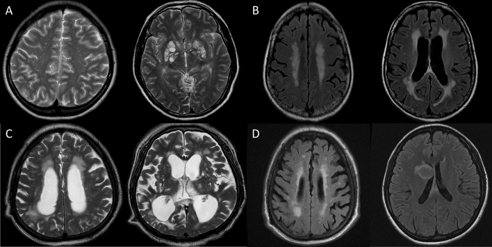
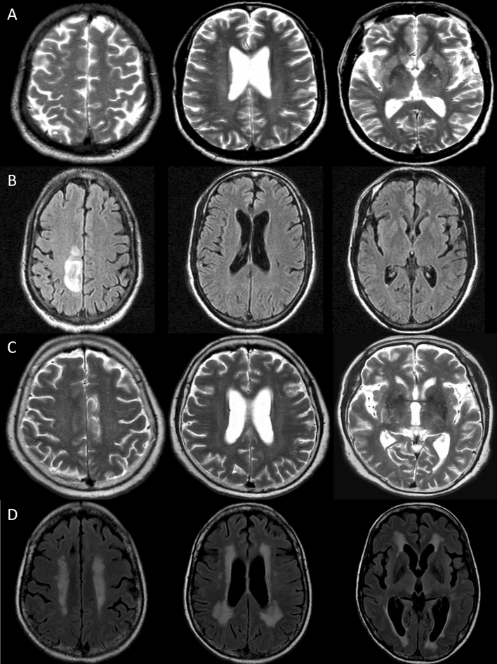
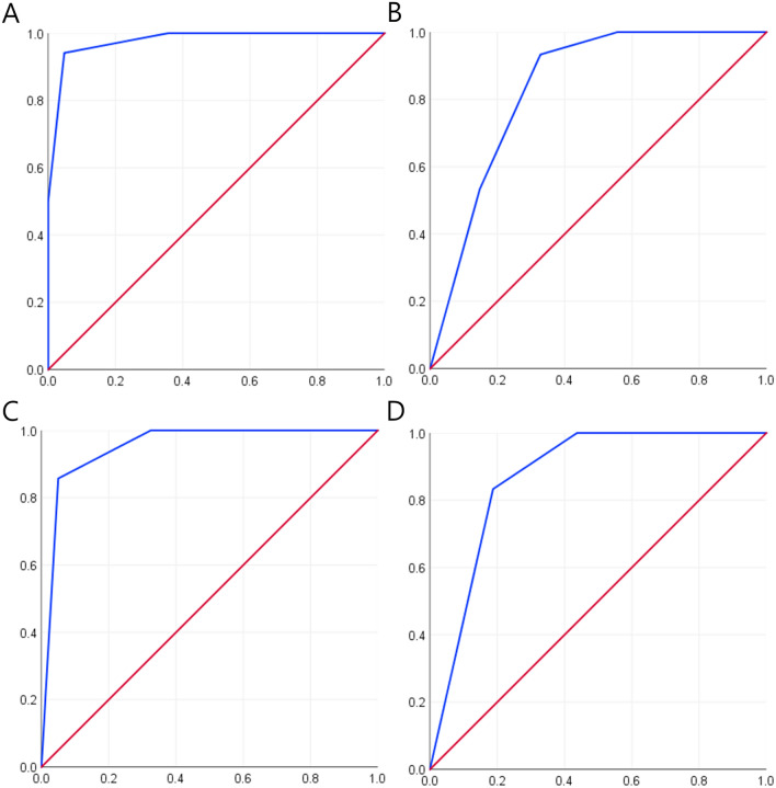
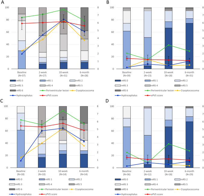
In Cryptococcus neoformans meningoencephalitis, brain MRI findings might reflect the phathomechanism of disease progression that is fungal accumulation in the peri-venular space and consequent invasion into the parenchyma. This study analyzed serial brain MRI findings of 76 patients with cryptococcus meningoencephalitis in association with the disease progression and outcomes. MRI parameters included the enlarged periventricular space (ePVS) score (range 0-8), periventricular lesion extension, cryptococcoma, and hydrocephalus. Clinical outcomes at 2-week, 10-week, and 6-month were evaluated using modified Rankin scale (mRS). At 6 months, 15 (19.7%) patients died and 34 (44.1%) had poor neurological outcomes (mRS scores > 2). At baseline, an ePVS score of ≥ 5 (Odds-ratio [OR]: 94.173, 95% confidence-interval [95%CI]: 7.507-1181.295, P < .001), periventricular lesion extension (OR: 51.965, 95%CI: 2.592-1041.673, P = .010), and presence of encephalitis feature (OR: 44.487, 95%CI: 1.689-1172.082, P = .023) were associated with 6-month poor outcomes. Presence of two or more risk factors among encephalitis feature, ePVS score ≥ 5, and periventricular lesion extension at baseline, was associated with 6-month poor outcomes (area under the curve [AUC]: 0.978, P < .001) and mortality (AUC: 0.836, P < .001). Disease progression was associated with interval development of cryptococcoma and hydrocephalus. Brain MRI findings might be useful in predicting outcomes and monitoring the progression of cryptococcus meningoencephalitis.
在新型隐球菌性脑膜脑炎中,脑 MRI 表现可能反映疾病进展的病理机制,即真菌在血管周围间隙蓄积并随之侵犯实质。本研究分析了 76 例新型隐球菌性脑膜脑炎患者的系列脑 MRI 表现与疾病进展和结局的关系。MRI 参数包括扩大的脑室周围间隙(ePVS)评分(范围 0-8)、脑室周围病变延伸、隐球菌肿和脑积水。使用改良 Rankin 量表(mRS)评估 2 周、10 周和 6 个月的临床结局。6 个月时,15 例(19.7%)患者死亡,34 例(44.1%)患者神经功能结局不良(mRS 评分>2)。基线时,ePVS 评分≥5(优势比[OR]:94.173,95%置信区间[95%CI]:7.507-1181.295,P<0.001)、脑室周围病变延伸(OR:51.965,95%CI:2.592-1041.673,P=0.010)和脑炎特征存在(OR:44.487,95%CI:1.689-1172.082,P=0.023)与 6 个月不良结局相关。基线时脑炎特征、ePVS 评分≥5 和脑室周围病变延伸中存在两个或更多危险因素与 6 个月不良结局(曲线下面积[AUC]:0.978,P<0.001)和死亡率(AUC:0.836,P<0.001)相关。疾病进展与隐球菌肿和脑积水的间隔性发展相关。脑 MRI 表现可能有助于预测结局和监测新型隐球菌性脑膜脑炎的进展。