Department of Neurosurgery, Li Hui Li Hospital of Medical Centre of Ningbo, No. 1111 Jiangnan Road, Yinzhou District, Ningbo, 315041, China.
Department of Internal Medicine, Ningbo Huamei Hospital University of Chinese Academy of Sciences, No. 41 Northwest Street, Ningbo, 315040, China.
BMC Surg. 2021 Mar 21;21(1):154. doi: 10.1186/s12893-021-01161-y.
Abdominal cerebrospinal fluid (CSF) pseudocyst is an uncommon but important complication of ventriculoperitoneal (VP) shunts. While individual articles have reported many cases of abdominal CSF pseudocyst following VP shunts, no case of a hemorrhagic abdominal pseudocyst after VP shunts has been reported so far.
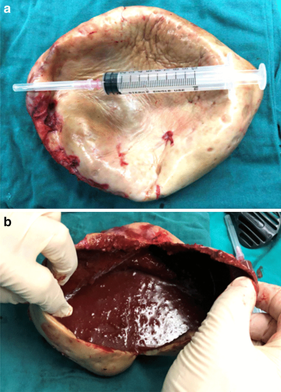
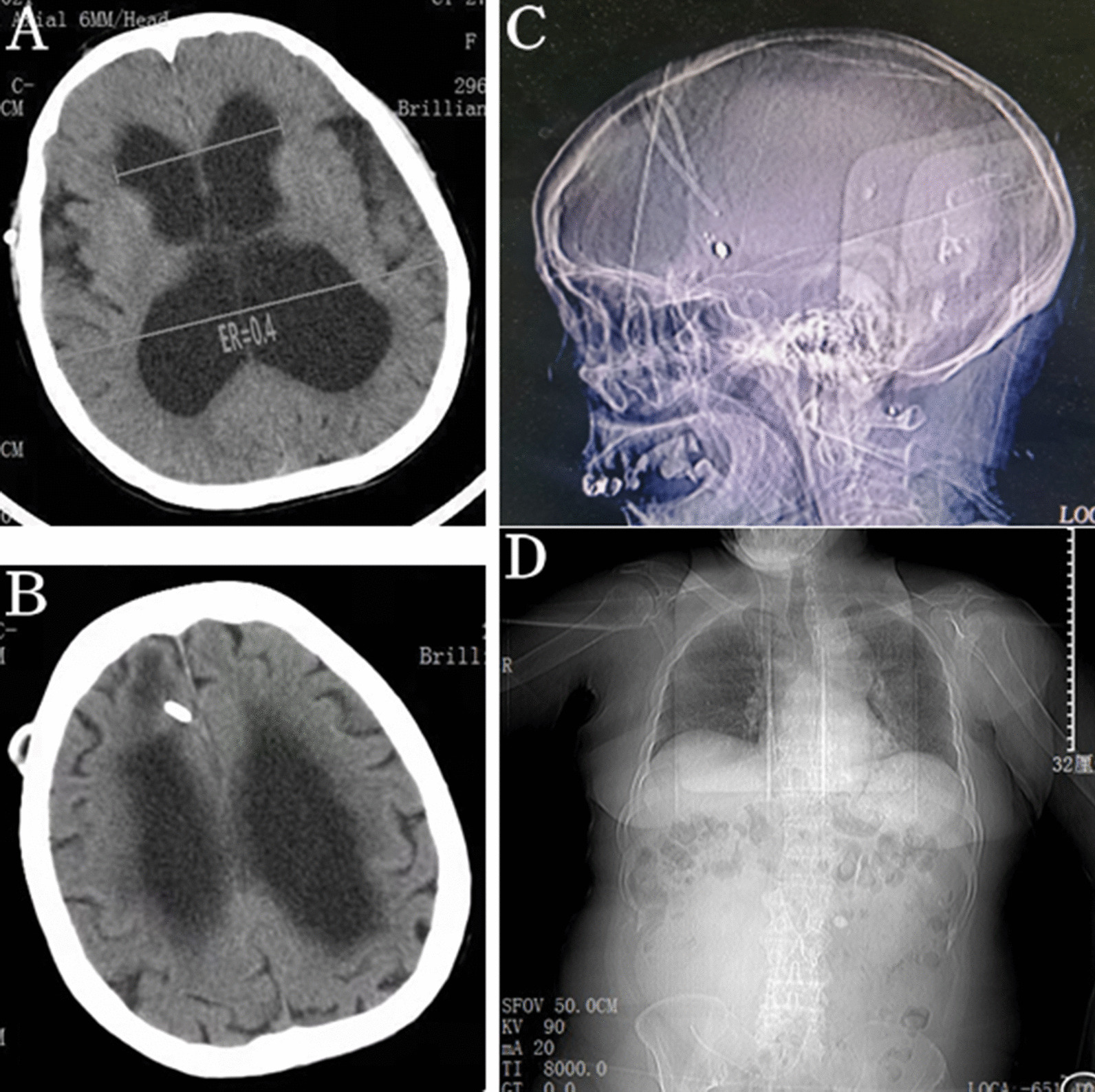
This article reports a 68-year-old woman with a 4-month history of progressive abdominal pain and distention. She denied any additional symptoms. A VP shunt was performed 15 years earlier to treat idiopathic normal pressure hydrocephalus and no other abdominal surgery was performed. Physical examination revealed an elastic palpable mass in her right lower abdomen, which was dull to percussion. Abdominal computed tomography (CT) scan indicated a large cystic collection of homogenous iso-density fluid in the right lower abdominal region with clear margins. The distal segment of the peritoneal shunt catheter was located within the cystic mass. Abdominal CSF pseudocyst was highly suspected as a diagnosis. Laparoscopic cyst drainage with removal of the whole cystic mass was performed, 15-cm cyst which found with thick walls and organized chronic hematic content. No responsible vessel for the cyst hemorrhage was identified. No further shunt revision was placed. Histological examination showed that the cyst wall consisted of outer fibrous tissue and inner granulation tissue without epithelial lining, and the cystic content was chronic hematoma. The patient had an uneventful postoperative course and remained asymptomatic for 8-mo follow-up.
To the best of our knowledge, this is the first report of hemorrhagic onset in the abdominal pseudocyst following VP shunt. Such special condition can accelerate the appearance of clinical signs of the abdominal pseudocyst after VP shunts, and its mechanisms may be similar to the evolution of subdural effusion into chronic subdural hematoma (CSDH).
腹部脑脊髓液(CSF)假性囊肿是脑室腹腔(VP)分流术后一种不常见但很重要的并发症。虽然个别文章报道了许多 VP 分流术后发生腹部 CSF 假性囊肿的病例,但迄今为止尚未报道过 VP 分流术后发生出血性腹部假性囊肿的病例。
本文报告了一例 68 岁女性,有 4 个月进行性腹痛和腹胀病史。她没有其他任何症状。15 年前,她因特发性正常压力脑积水行 VP 分流术,此后未行任何其他腹部手术。体格检查发现其右下腹部有一可触及的弹性肿块,叩诊呈浊音。腹部 CT 扫描显示右下腹部有一个大的囊性均匀等密度液体积聚,边界清晰。腹膜分流管的远端位于囊性肿块内。高度怀疑诊断为腹部 CSF 假性囊肿。行腹腔镜囊肿引流术,切除整个囊肿肿块,发现 15cm 厚壁、有组织的慢性血性内容物的囊肿。未发现导致囊肿出血的责任血管。未再放置分流管。组织学检查显示囊肿壁由外纤维组织和内肉芽组织组成,无上皮衬里,囊内容物为慢性血肿。患者术后恢复顺利,无并发症,随访 8 个月无症状。
据我们所知,这是首例报道 VP 分流术后腹部假性囊肿出血的病例。这种特殊情况可能会加速 VP 分流术后腹部假性囊肿出现临床症状,其机制可能与硬膜下积液演变为慢性硬膜下血肿(CSDH)相似。