• 文献检索
  • 文档翻译
  • 深度研究
  • 学术资讯
  • Suppr Zotero 插件Zotero 插件
  • 邀请有礼
  • 套餐&价格
  • 历史记录
应用&插件
Suppr Zotero 插件Zotero 插件浏览器插件Mac 客户端Windows 客户端微信小程序
定价
高级版会员购买积分包购买API积分包
服务
文献检索文档翻译深度研究API 文档MCP 服务
关于我们
关于 Suppr公司介绍联系我们用户协议隐私条款
关注我们

Suppr 超能文献

核心技术专利:CN118964589B侵权必究
粤ICP备2023148730 号-1Suppr @ 2026

文献检索

告别复杂PubMed语法,用中文像聊天一样搜索,搜遍4000万医学文献。AI智能推荐,让科研检索更轻松。

立即免费搜索

文件翻译

保留排版,准确专业,支持PDF/Word/PPT等文件格式,支持 12+语言互译。

免费翻译文档

深度研究

AI帮你快速写综述,25分钟生成高质量综述,智能提取关键信息,辅助科研写作。

立即免费体验

大脑 Gali 静脉周围深部静脉的 CT 血管造影研究。

CT angiographic study of the cerebral deep veins around the vein of Galen.

机构信息

Department of Neurosurgery, The First Hospital of Jilin University, Changchun, 130021, China.

Department of Radiology, The First Hospital of Jilin University, Changchun, 130021, China.

出版信息

Int J Med Sci. 2021 Feb 6;18(7):1699-1710. doi: 10.7150/ijms.54891. eCollection 2021.

DOI:10.7150/ijms.54891
PMID:33746586
原文链接:https://pmc.ncbi.nlm.nih.gov/articles/PMC7976576/
Abstract

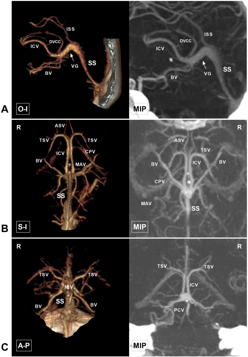
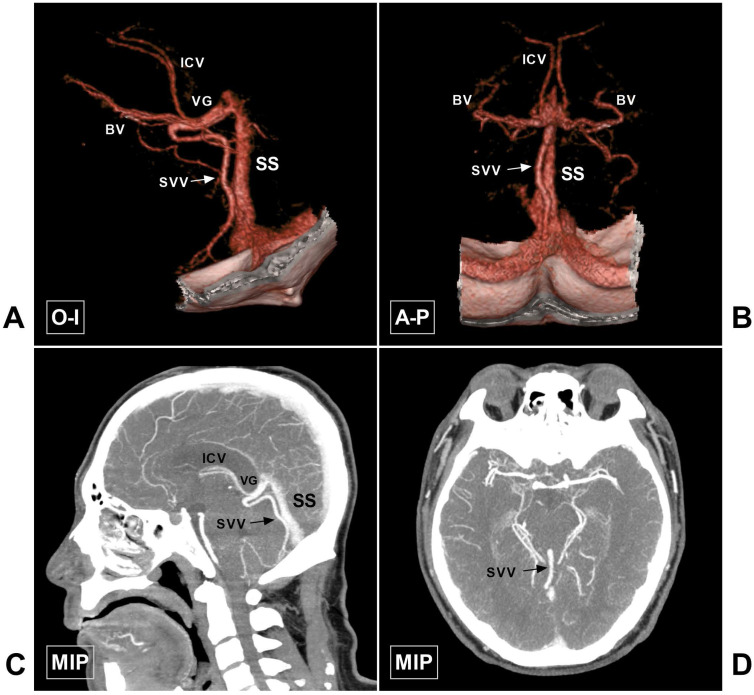
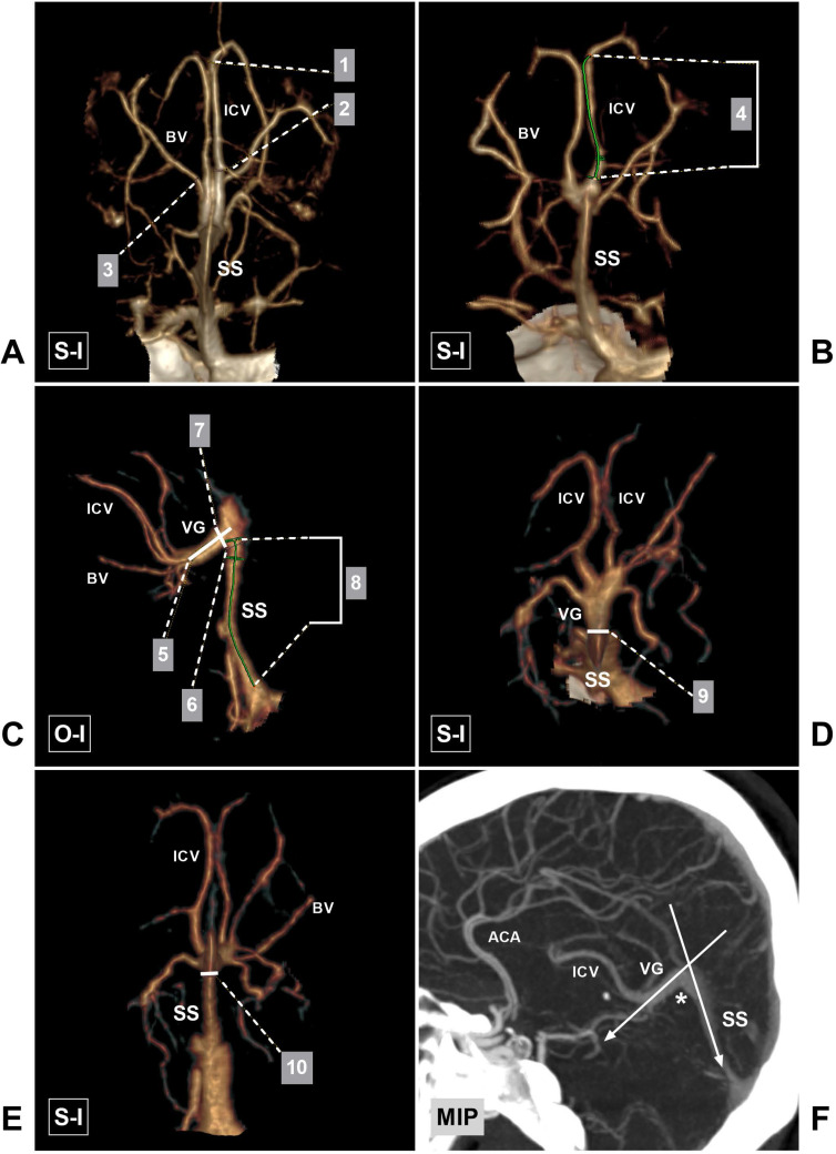
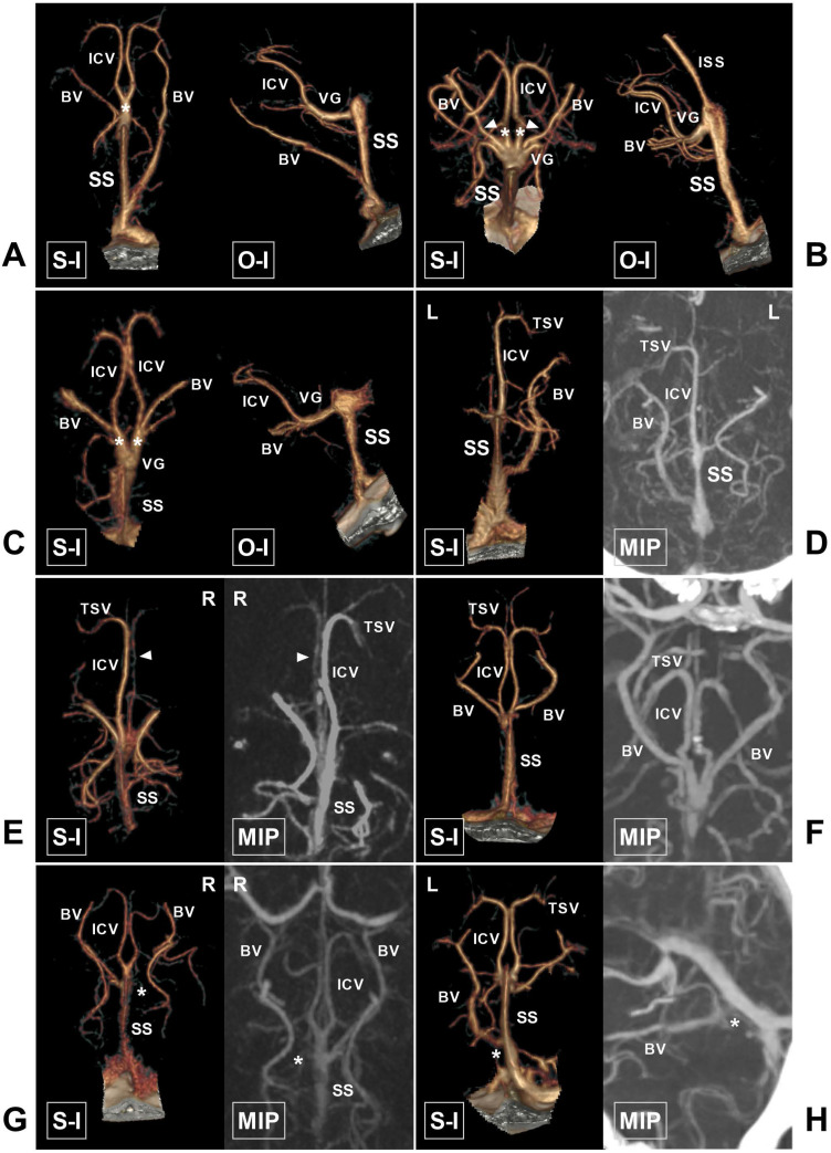
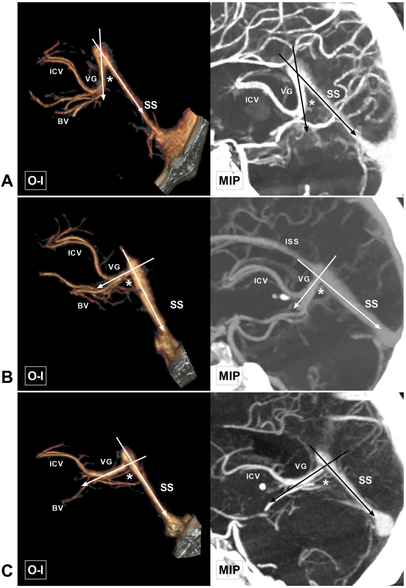
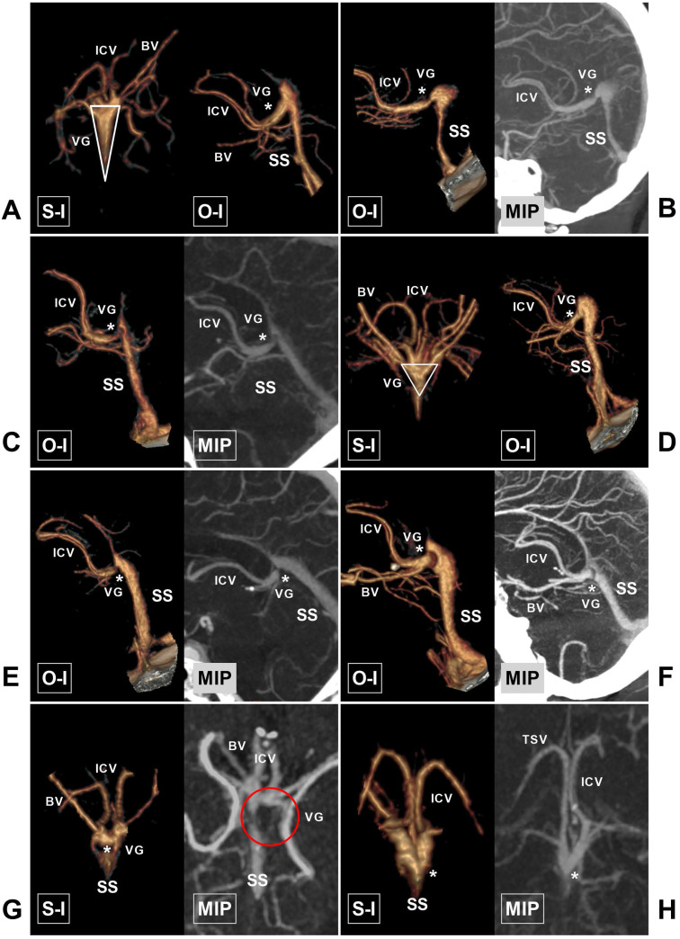
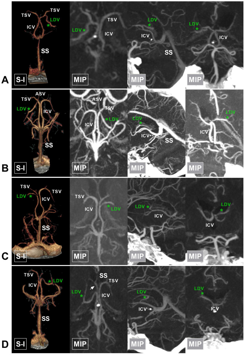
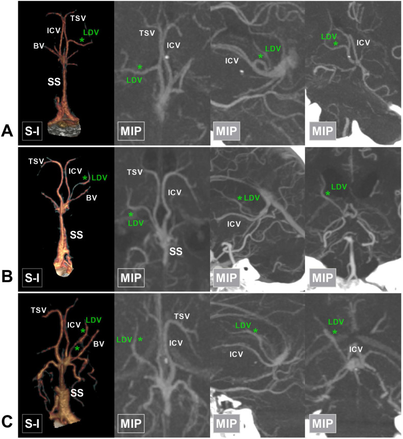
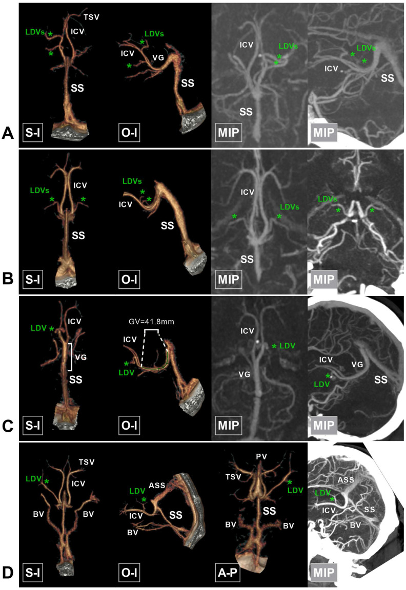
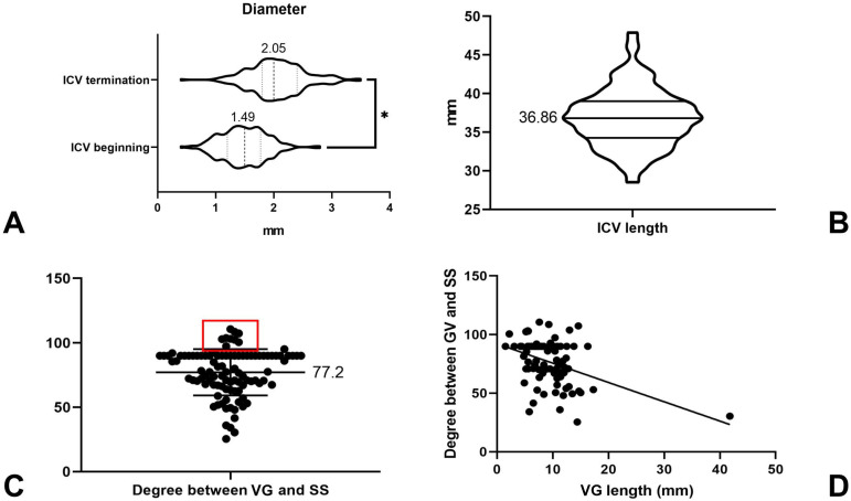
Research on the anatomy of cerebral deep veins (CDVs) around the vein of Galen (VG) is very important and has fundamental clinical significance. Large-scale anatomical studies of CDVs using computed tomography angiography (CTA) are rarely reported. A retrospective study of the CDVs around the VG was conducted in Chinese patients of Han nationality. One hundred cases were included in the final analysis. The patients were aged from 17 to 78 years (mean: 42.3 years). Also, 46% of the patients were female. The diameter of the internal cerebral vein (ICV) at its beginning and termination points ranged from 0.4 to 2.8 mm (1.49 ± 0.39 mm) and 0.4 to 3.5 mm (2.05 ± 0.47 mm), respectively. There was statistical significance regarding the diameter of the ICV at its beginning and termination points (P <0.01). The ICV length ranged from 28.5 to 47.9 mm (36.86 ± 3.74 mm). The length of the straight sinus (SS) ranged from 30.2 to 57.8 mm (43.6 ± 6.37 mm). The length of the VG ranged from 1.5 to 41.8 mm (9.30 ± 4.76 mm). The angle at the VG and SS transition area ranged from 25.4 to 110.6° (77.2 ± 18.0°). This study was a meaningful attempt to conduct anatomical research of CDVs using CTA. Preoperative familiarity with the normal venous structure and its variation around the VG would be helpful for endovascular treatment.

摘要

关于大脑深静脉(CDVs)在Galen 静脉(VG)周围的解剖学研究非常重要,具有重要的临床意义。使用计算机断层血管造影术(CTA)对 CDVs 进行大规模解剖研究很少有报道。本研究对汉族中国患者的 VG 周围 CDVs 进行了回顾性研究。最终纳入 100 例患者进行分析。患者年龄 17-78 岁(平均年龄:42.3 岁),其中 46%为女性。内脑静脉(ICV)起始点和终点处的直径范围为 0.4-2.8mm(1.49±0.39mm)和 0.4-3.5mm(2.05±0.47mm),差异有统计学意义(P<0.01)。ICV 长度范围为 28.5-47.9mm(36.86±3.74mm)。直窦(SS)长度范围为 30.2-57.8mm(43.6±6.37mm)。VG 长度范围为 1.5-41.8mm(9.30±4.76mm)。VG 和 SS 过渡区的角度范围为 25.4-110.6°(77.2±18.0°)。本研究是使用 CTA 对 CDVs 进行解剖学研究的一次有意义的尝试。术前熟悉 VG 周围正常静脉结构及其变异,有助于血管内治疗。

https://cdn.ncbi.nlm.nih.gov/pmc/blobs/aee1/7976576/5e788cc4269d/ijmsv18p1699g010.jpg
https://cdn.ncbi.nlm.nih.gov/pmc/blobs/aee1/7976576/c559a8ff3d98/ijmsv18p1699g001.jpg
https://cdn.ncbi.nlm.nih.gov/pmc/blobs/aee1/7976576/8f4313266219/ijmsv18p1699g002.jpg
https://cdn.ncbi.nlm.nih.gov/pmc/blobs/aee1/7976576/5efd8f06a6d0/ijmsv18p1699g003.jpg
https://cdn.ncbi.nlm.nih.gov/pmc/blobs/aee1/7976576/2ef674ebcef4/ijmsv18p1699g004.jpg
https://cdn.ncbi.nlm.nih.gov/pmc/blobs/aee1/7976576/4194618653d6/ijmsv18p1699g005.jpg
https://cdn.ncbi.nlm.nih.gov/pmc/blobs/aee1/7976576/878290ab8611/ijmsv18p1699g006.jpg
https://cdn.ncbi.nlm.nih.gov/pmc/blobs/aee1/7976576/7858bfb9c5f6/ijmsv18p1699g007.jpg
https://cdn.ncbi.nlm.nih.gov/pmc/blobs/aee1/7976576/3ef99e10eb41/ijmsv18p1699g008.jpg
https://cdn.ncbi.nlm.nih.gov/pmc/blobs/aee1/7976576/363bf65e8a6d/ijmsv18p1699g009.jpg
https://cdn.ncbi.nlm.nih.gov/pmc/blobs/aee1/7976576/5e788cc4269d/ijmsv18p1699g010.jpg

相似文献

1
CT angiographic study of the cerebral deep veins around the vein of Galen.大脑 Gali 静脉周围深部静脉的 CT 血管造影研究。
Int J Med Sci. 2021 Feb 6;18(7):1699-1710. doi: 10.7150/ijms.54891. eCollection 2021.
2
Three-dimensional computed tomography angiography of the galenic system for the occipital transtentorial approach.用于枕下经小脑幕入路的大脑大静脉系统三维计算机断层血管造影
Neurol Med Chir (Tokyo). 2005 Aug;45(8):387-93; discussion 393-4. doi: 10.2176/nmc.45.387.
3
[CT angiography of cerebral venous circulation: anatomical visualization and diagnostic pitfalls in interpretation].[脑静脉循环的CT血管造影:解剖可视化及解读中的诊断陷阱]
Rofo. 1997 Dec;167(6):612-8. doi: 10.1055/s-2007-1015591.
4
The Anatomy of the Convergence of Major Deep Cerebral Veins in the Pineal Region: A Computed Tomography Angiography Study.松果体区大脑大静脉汇合部的解剖结构:一项 CT 血管造影研究。
World Neurosurg. 2021 Mar;147:e334-e342. doi: 10.1016/j.wneu.2020.12.057. Epub 2020 Dec 17.
5
Cranial venous sinus dominance: what to expect? Analysis of 100 cerebral angiographies.颅内静脉窦优势:预期结果如何?对100例脑血管造影的分析。
Arq Neuropsiquiatr. 2017 May;75(5):295-300. doi: 10.1590/0004-282X20170042.
6
Anatomy of cerebral veins and sinuses.脑静脉和静脉窦的解剖结构。
Front Neurol Neurosci. 2008;23:4-15. doi: 10.1159/000111256.
7
Recovery from primary deep cerebral venous sinus thrombosis with recanalisation.原发性大脑静脉窦血栓形成伴再通后的恢复情况。
Neuroradiology. 1995 Nov;37(8):645-8. doi: 10.1007/BF00593380.
8
[Collateral venous channels in occlusion of deep cerebral veins and sinuses].[大脑深部静脉和静脉窦闭塞时的侧支静脉通道]
No To Shinkei. 1994 Oct;46(10):935-9.
9
Anatomy of the deep venous system in vein of Galen malformation and its changes after endovascular treatment depicted by magnetic resonance venography.磁共振静脉造影显示 Galién 静脉畸形深部静脉系统的解剖结构及其血管内治疗后的变化。
J Neurointerv Surg. 2019 Jan;11(1):84-89. doi: 10.1136/neurintsurg-2018-013789. Epub 2018 May 24.
10
Deep venous communication in vein of Galen malformations: incidence, Imaging, and Implications for treatment.Galen 静脉畸形中的深静脉交通:发生率、影像学表现及对治疗的影响。
J Neurointerv Surg. 2021 Mar;13(3):290-293. doi: 10.1136/neurintsurg-2020-016224. Epub 2020 Jun 16.

引用本文的文献

1
Effect of moyamoya disease on the basilar artery and adjacent arteries on CTA.烟雾病对CTA上基底动脉及相邻动脉的影响。
eNeurologicalSci. 2024 May 5;35:100501. doi: 10.1016/j.ensci.2024.100501. eCollection 2024 Jun.
2
Clinical outcome and prognostic factors of patients with non-traumatic angiography-negative subarachnoid hemorrhage.非创伤性血管造影阴性蛛网膜下腔出血患者的临床结局及预后因素
Front Neurol. 2023 Jul 20;14:1157845. doi: 10.3389/fneur.2023.1157845. eCollection 2023.
3
CTA Study of Ruptured Aneurysms of the Posterior Communicating Artery.

本文引用的文献

1
Anatomical features of the superficial temporal artery in hemorrhagic moyamoya disease based on CT angiography.基于CT血管造影的出血型烟雾病颞浅动脉的解剖学特征
Exp Ther Med. 2020 Mar;19(3):2143-2148. doi: 10.3892/etm.2020.8473. Epub 2020 Jan 27.
2
The Internal Cerebral Vein: New Classification of Branching Patterns Based on CTA.颅内静脉:基于 CTA 的新分支模式分类。
AJNR Am J Neuroradiol. 2019 Oct;40(10):1719-1724. doi: 10.3174/ajnr.A6200. Epub 2019 Sep 5.
3
Transvenous embolization of brain arteriovenous malformations: a review of techniques, indications, and outcomes.
后交通动脉破裂动脉瘤的CT血管造影研究
Stroke Res Treat. 2022 Sep 14;2022:5774735. doi: 10.1155/2022/5774735. eCollection 2022.
4
Current status of perimesencephalic non-aneurysmal subarachnoid hemorrhage.中脑周围非动脉瘤性蛛网膜下腔出血的现状
Front Neurol. 2022 Sep 1;13:960702. doi: 10.3389/fneur.2022.960702. eCollection 2022.
5
Role of the anterior choroidal artery in the endovascular treatment of brain arteriovenous malformations.脉络膜前动脉在脑动静脉畸形血管内治疗中的作用。
Acta Neurol Belg. 2023 Feb;123(1):57-67. doi: 10.1007/s13760-022-01878-4. Epub 2022 Feb 11.
经静脉脑动静脉畸形栓塞术:技术、适应证和结果的综述。
Neurosurg Focus. 2018 Jul;45(1):E13. doi: 10.3171/2018.3.FOCUS18113.
4
Microsurgical anatomy of the central core of the brain.脑中央核的显微解剖
J Neurosurg. 2018 Sep;129(3):752-769. doi: 10.3171/2017.5.JNS162897. Epub 2017 Dec 22.
5
Encountering a Basal Vein of Rosenthal Variant During the Anterior Transpetrosal Approach.经岩前入路中遇到罗森塔尔基底静脉变异
World Neurosurg. 2016 Feb;86:513.e19-22. doi: 10.1016/j.wneu.2015.09.030. Epub 2015 Sep 25.
6
Anatomical variants of the basal vein of Rosenthal: prevalence in idiopathic subarachnoid hemorrhage.罗森塔尔基底静脉的解剖变异:在特发性蛛网膜下腔出血中的发生率
Acta Neurochir (Wien). 2014 Jan;156(1):45-51. doi: 10.1007/s00701-013-1907-6. Epub 2013 Oct 18.
7
Anatomy of cerebral veins and sinuses.脑静脉和静脉窦的解剖结构。
Front Neurol Neurosci. 2008;23:4-15. doi: 10.1159/000111256.
8
Surgical anatomy and landmarks for the basal vein of rosenthal.罗森塔尔基底静脉的手术解剖及标志
J Neurosurg. 2007 May;106(5):900-2. doi: 10.3171/jns.2007.106.5.900.
9
Microsurgical anatomy of the great cerebral vein of Galen and its tributaries.大脑大静脉(Galen静脉)及其属支的显微外科解剖
J Neurosurg. 2003 Dec;99(6):1028-38. doi: 10.3171/jns.2003.99.6.1028.
10
Perimesencephalic nonaneurysmal subarachnoid haemorrhage and variations in the veins.中脑周围非动脉瘤性蛛网膜下腔出血与静脉变异
Neuroradiology. 2002 Apr;44(4):319-25. doi: 10.1007/s00234-001-0741-3. Epub 2002 Feb 2.