Soattin Luca, Borbas Zoltan, Caldwell Jane, Prendergast Brian, Vohra Akbar, Saeed Yawer, Hoschtitzky Andreas, Yanni Joseph, Atkinson Andrew, Logantha Sunil Jit, Borbas Balint, Garratt Clifford, Morris Gwilym Matthew, Dobrzynski Halina
Division of Cardiovascular Sciences, Faculty of Biology, Medicine and Health, Manchester Academic Health Science Centre, University of Manchester, Manchester, United Kingdom.
Manchester Heart Centre, Central Manchester University Foundation Trust, Manchester Academic Health Science Centre, Manchester, United Kingdom.
Front Physiol. 2021 Mar 4;12:592229. doi: 10.3389/fphys.2021.592229. eCollection 2021.
The sinoatrial/sinus node (SAN) is the primary pacemaker of the heart. In humans, SAN is surrounded by the paranodal area (PNA). Although the PNA function remains debated, it is thought to act as a subsidiary atrial pacemaker (SAP) tissue and become the dominant pacemaker in the setting of sinus node disease (SND). Large animal models of SND allow characterization of SAP, which might be a target for novel treatment strategies for SAN diseases.
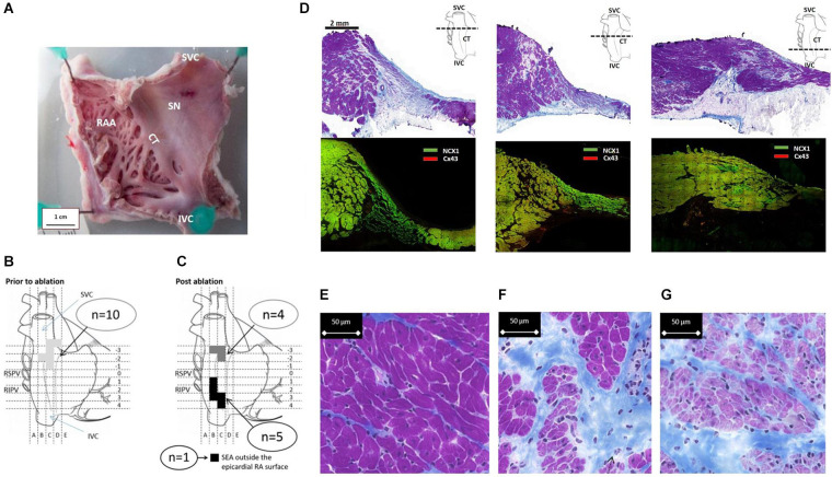
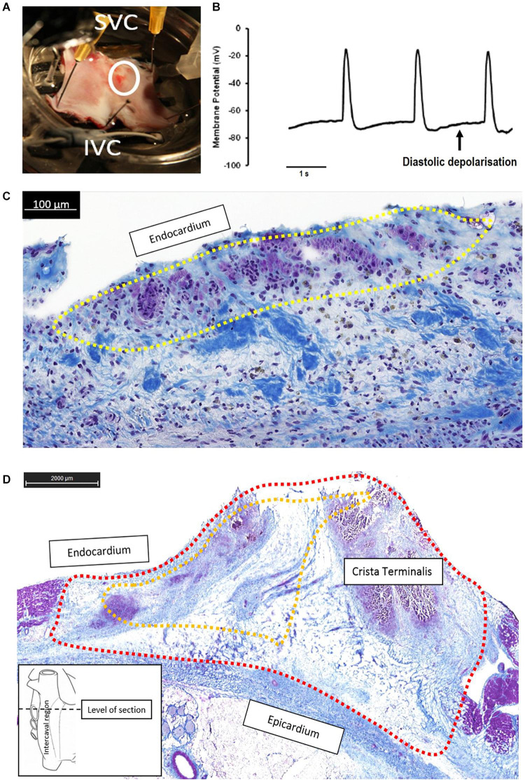
A goat model of SND was developed ( = 10) by epicardially ablating the SAN and validated by mapping of emergent SAP locations through an ablation catheter and surface electrocardiogram (ECG). Structural characterization of the goat SAN and SAP was assessed by histology and immunofluorescence techniques.
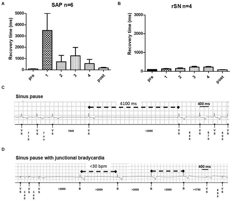
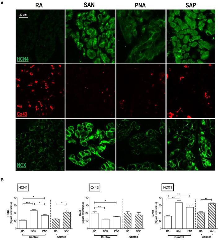
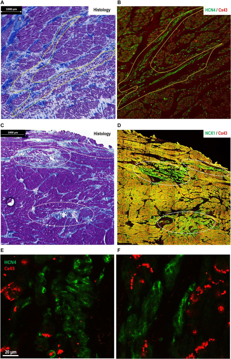
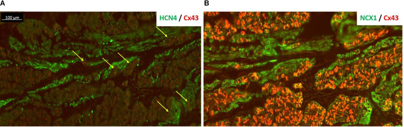
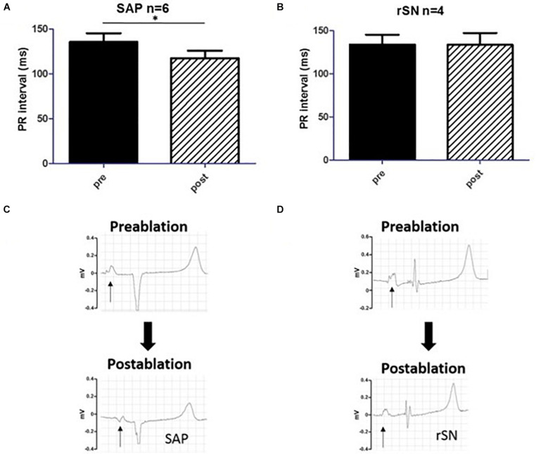
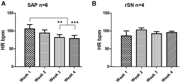
When the SAN was ablated, SAPs featured a shortened atrioventricular conduction, consistent with the location in proximity of atrioventricular junction. SAP recovery time showed significant prolongation compared to the SAN recovery time, followed by a decrease over a follow-up of 4 weeks. Like the SAN tissue, the SAP expressed the main isoform of pacemaker hyperpolarization-activated cyclic nucleotide-gated channel 4 (HCN4) and Na/Ca exchanger 1 (NCX1) and no high conductance connexin 43 (Cx43). Structural characterization of the right atrium (RA) revealed that the SAN was located at the earliest activation [i.e., at the junction of the superior vena cava (SVC) with the RA] and was surrounded by the paranodal-like tissue, extending down to the inferior vena cava (IVC). Emerged SAPs were localized close to the IVC and within the thick band of the atrial muscle known as the crista terminalis (CT).
SAN ablation resulted in the generation of chronic SAP activity in 60% of treated animals. SAP displayed development over time and was located within the previously discovered PNA in humans, suggesting its role as dominant pacemaker in SND. Therefore, SAP in goat constitutes a promising stable target for electrophysiological modification to construct a fully functioning pacemaker.
窦房结(SAN)是心脏的主要起搏点。在人类中,窦房结被结旁区域(PNA)包围。尽管结旁区域的功能仍存在争议,但人们认为它可作为心房辅助起搏点(SAP)组织,并在窦房结疾病(SND)情况下成为主导起搏点。大型动物的窦房结疾病模型有助于对辅助起搏点进行特征描述,这可能是窦房结疾病新型治疗策略的靶点。
通过心外膜消融窦房结建立了山羊窦房结疾病模型(n = 10),并通过消融导管和体表心电图(ECG)对新出现的辅助起搏点位置进行标测来验证。通过组织学和免疫荧光技术评估山羊窦房结和辅助起搏点的结构特征。
当窦房结被消融时,辅助起搏点的房室传导缩短,这与房室交界附近的位置一致。与窦房结恢复时间相比,辅助起搏点恢复时间显著延长,随后在4周的随访中有所缩短。与窦房结组织一样,辅助起搏点表达起搏超极化激活环核苷酸门控通道4(HCN4)和钠钙交换体1(NCX1)的主要异构体,且不表达高电导连接蛋白43(Cx43)。右心房(RA)的结构特征显示,窦房结位于最早激活处[即上腔静脉(SVC)与右心房的交界处],并被结旁样组织包围,向下延伸至下腔静脉(IVC)。新出现的辅助起搏点位于下腔静脉附近以及心房肌的厚带即界嵴(CT)内。
窦房结消融导致60%的受试动物产生慢性辅助起搏点活动。辅助起搏点随时间发展,并位于人类先前发现的结旁区域内,表明其在窦房结疾病中作为主导起搏点的作用。因此,山羊的辅助起搏点是构建功能完备起搏器的电生理改造的一个有前景的稳定靶点。