van Zanden Judith E, Leuvenink Henri G D, Verschuuren Erik A M, Veldhuis Zwanida J, Ottens Petra J, Erasmus Michiel E, Hottenrott Maximilia C
Department of Surgery, University Medical Center Groningen, Groningen, The Netherlands.
Department of Pulmonary Diseases, University Medical Center Groningen, Groningen, The Netherlands.
Transplant Direct. 2021 Mar 16;7(4):e682. doi: 10.1097/TXD.0000000000001141. eCollection 2021 Apr.
The onset of brain death (BD) leads to the deterioration of potential donor lungs. Methylprednisolone is considered to increase lung oxygenation capacity and enhance the procurement yield of donor lungs, when applied in situ, during donor management. However, whether BD-induced lung damage is ameliorated upon treatment with methylprednisolone during acellular ex vivo lung perfusion (EVLP), remains unknown. We aimed to investigate whether the quality of lungs from brain-dead donors improves upon methylprednisolone treatment during EVLP.
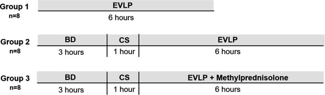
Rat lungs were randomly assigned to 1 of 3 experimental groups (n = 8/group): (1) healthy, directly procured lungs subjected to EVLP; (2) lungs from brain-dead rats subjected to cold storage and EVLP; and (3) lungs from brain-dead rats subjected to cold storage and EVLP with 40 mg methylprednisolone added to the perfusate. Ventilation and perfusion parameters, histology, edema formation, metabolic profile, and inflammatory status of lungs were investigated.
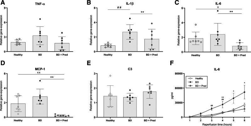
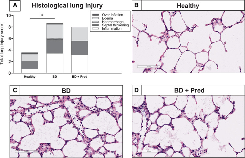
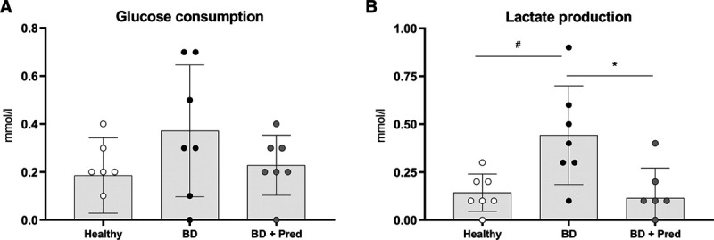
Methylprednisolone treated lungs from brain-dead donors improved positive inspiratory pressures needed to maintain tidal volumes of 7 mL/kg of body weight, which was 25.6 ± 5.8 cm HO in untreated lungs and 18.0 ± 3.0 cm HO in methylprednisolone treated lungs, after 6 h EVLP. Furthermore, dynamic lung compliance increased upon methylprednisolone treatment, with values of 0.11 ± 0.05 mL/cm HO versus 0.18 ± 0.04 mL/cm HO after 6 h of EVLP. Methylprednisolone treatment ameliorated the amount of lung edema, as corroborated by a reduction of 0.7 in the wet/dry ratio. Although glucose consumption levels were comparable, the BD-induced cumulative lactate production decreased from 0.44 ± 0.26 to 0.11 ± 0.16 mmol/L upon methylprednisolone treatment. Finally, BD-induced inflammatory status was reduced upon methylprednisolone treatment compared to untreated lungs from brain-dead donors, as reflected by lower proinflammatory gene expression levels of IL-1β, IL-6 and MCP-1, and IL-6 perfusate levels.
We showed that methylprednisolone treatment during EVLP attenuates BD-induced lung injury.
脑死亡(BD)的发生会导致潜在供体肺功能恶化。在供体管理过程中,甲基强的松龙在原位应用时,被认为可提高肺的氧合能力并提高供体肺的获取率。然而,在脱细胞体外肺灌注(EVLP)期间,用甲基强的松龙治疗是否能改善BD诱导的肺损伤,仍不清楚。我们旨在研究在EVLP期间,甲基强的松龙治疗是否能改善脑死亡供体肺的质量。
将大鼠肺随机分为3个实验组之一(每组n = 8):(1)健康的、直接获取的肺进行EVLP;(2)来自脑死亡大鼠的肺进行冷藏并进行EVLP;(3)来自脑死亡大鼠的肺进行冷藏并进行EVLP,在灌注液中添加40mg甲基强的松龙。研究了肺的通气和灌注参数、组织学、水肿形成、代谢谱和炎症状态。
在EVLP 6小时后,甲基强的松龙治疗的脑死亡供体肺改善了维持7mL/kg体重潮气量所需的正吸气压力,未治疗的肺为25.6±5.8cm H₂O,甲基强的松龙治疗的肺为18.0±3.0cm H₂O。此外,甲基强的松龙治疗后动态肺顺应性增加,EVLP 6小时后的值为0.11±0.05mL/cm H₂O,而未治疗的为0.18±0.04mL/cm H₂O。甲基强的松龙治疗改善了肺水肿的程度,湿/干比降低了0.7证实了这一点。尽管葡萄糖消耗水平相当,但甲基强的松龙治疗后,BD诱导的累积乳酸产量从0.44±0.26降至0.11±0.16mmol/L。最后,与未治疗的脑死亡供体肺相比,甲基强的松龙治疗降低了BD诱导的炎症状态,这通过IL-1β、IL-6和MCP-1的促炎基因表达水平降低以及IL-6灌注液水平反映出来。
我们表明,在EVLP期间,甲基强的松龙治疗可减轻BD诱导的肺损伤。