Department of Medicine, Icahn School of Medicine at Mount Sinai, Mount Sinai Beth Israel, First Avenue, 16th Street, New York City, NY, 10003, USA.
Division of Pulmonary Medicine, Department of Medicine, Keio University School of Medicine, Tokyo, Japan.
BMC Pulm Med. 2021 Mar 22;21(1):97. doi: 10.1186/s12890-021-01463-0.
The coronavirus disease 2019 (COVID-19) causes a wide spectrum of lung manifestations ranging from mild asymptomatic disease to severe respiratory failure. We aimed to clarify the characteristics of radiological and functional lung sequelae of COVID-19 patients described in follow-up period.
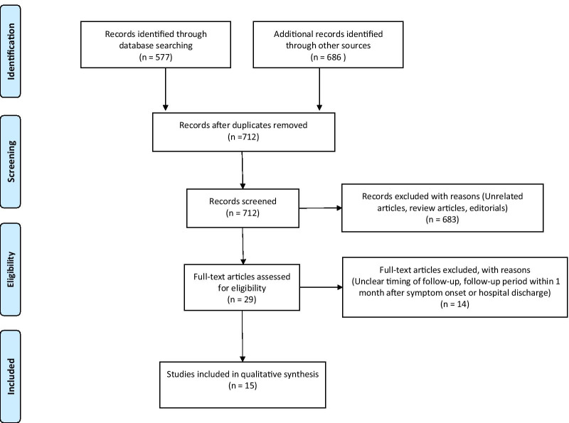
PubMed and EMBASE were searched on January 20th, 2021 to investigate characteristics of lung sequelae in COVID-19 patients. Chest computed tomography (CT) and pulmonary function test (PFT) data were collected and analyzed using one-group meta-analysis.
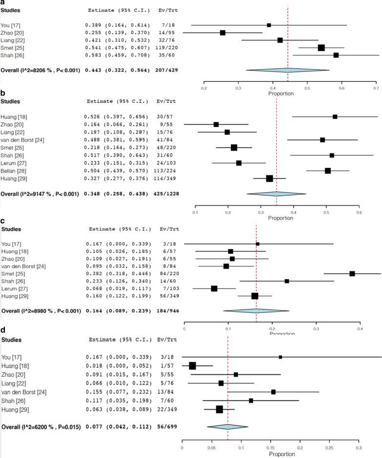
Our search identified 15 eligible studies with follow-up period in a range of 1-6 months. A total of 3066 discharged patients were included in these studies. Among them, 1232 and 1359 patients were evaluated by chest CT and PFT, respectively. The approximate follow-up timing on average was 90 days after either symptom onset or hospital discharge. The frequency of residual CT abnormalities after hospital discharge was 55.7% (95% confidential interval (CI) 41.2-70.1, I = 96.2%). The most frequent chest CT abnormality was ground glass opacity in 44.1% (95% CI 30.5-57.8, I = 96.2%), followed by parenchymal band or fibrous stripe in 33.9% (95% CI 18.4-49.4, I = 95.0%). The frequency of abnormal pulmonary function test was 44.3% (95% CI 32.2-56.4, I = 82.1%), and impaired diffusion capacity was the most frequently observed finding in 34.8% (95% CI 25.8-43.8, I = 91.5%). Restrictive and obstructive patterns were observed in 16.4% (95% CI 8.9-23.9, I = 89.8%) and 7.7% (95% CI 4.2-11.2, I = 62.0%), respectively.
This systematic review suggested that about half of the patients with COVID-19 still had residual abnormalities on chest CT and PFT at about 3 months. Further studies with longer follow-up term are warranted.
2019 年冠状病毒病(COVID-19)可引起广泛的肺部表现,从轻症无症状疾病到严重呼吸衰竭不等。我们旨在阐明 COVID-19 患者在随访期间描述的放射学和功能肺部后遗症的特征。
于 2021 年 1 月 20 日在 PubMed 和 EMBASE 上进行搜索,以调查 COVID-19 患者肺部后遗症的特征。使用单组荟萃分析收集和分析胸部计算机断层扫描(CT)和肺功能检查(PFT)数据。
我们的搜索确定了 15 项符合条件的研究,随访时间范围为 1-6 个月。这些研究共纳入 3066 名出院患者,其中 1232 名和 1359 名患者分别接受了胸部 CT 和 PFT 评估。平均随访时间平均为症状发作或出院后 90 天。出院后残留 CT 异常的频率为 55.7%(95%可信区间[CI] 41.2-70.1,I=96.2%)。最常见的胸部 CT 异常为磨玻璃影占 44.1%(95%CI 30.5-57.8,I=96.2%),其次为实质带或纤维条纹占 33.9%(95%CI 18.4-49.4,I=95.0%)。肺功能检查异常的频率为 44.3%(95%CI 32.2-56.4,I=82.1%),最常见的是弥散功能受损占 34.8%(95%CI 25.8-43.8,I=91.5%)。限制性和阻塞性模式分别占 16.4%(95%CI 8.9-23.9,I=89.8%)和 7.7%(95%CI 4.2-11.2,I=62.0%)。
本系统评价表明,大约一半的 COVID-19 患者在大约 3 个月时胸部 CT 和 PFT 仍存在残留异常。需要进一步进行随访时间更长的研究。