Englander Zachary K, Wei Hong-Jian, Pouliopoulos Antonios N, Bendau Ethan, Upadhyayula Pavan, Jan Chia-Ing, Spinazzi Eleanora F, Yoh Nina, Tazhibi Masih, McQuillan Nicholas M, Wang Tony J C, Bruce Jeffrey N, Canoll Peter, Feldstein Neil A, Zacharoulis Stergios, Konofagou Elisa E, Wu Cheng-Chia
Department of Neurological Surgery, Columbia University Irving Medical Center, New York, NY, 10032, USA.
Department of Radiation Oncology, Columbia University Irving Medical Center, 622 W. 168th Street, New York, NY, 10032, USA.
Sci Rep. 2021 Mar 22;11(1):6521. doi: 10.1038/s41598-021-85180-y.
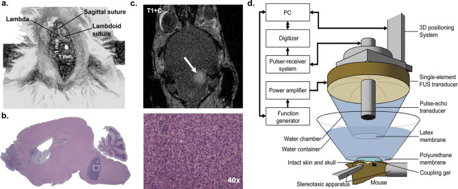
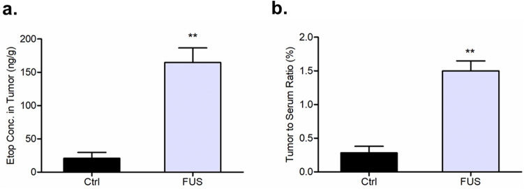
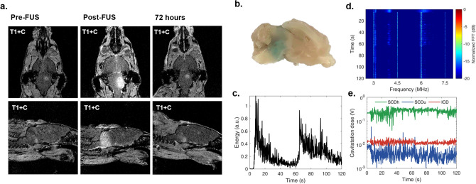
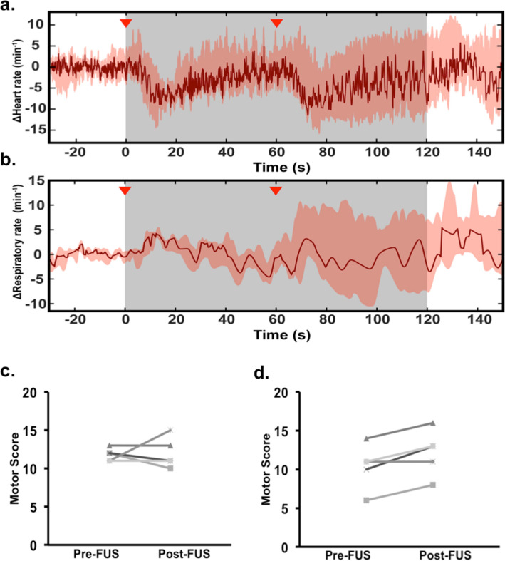
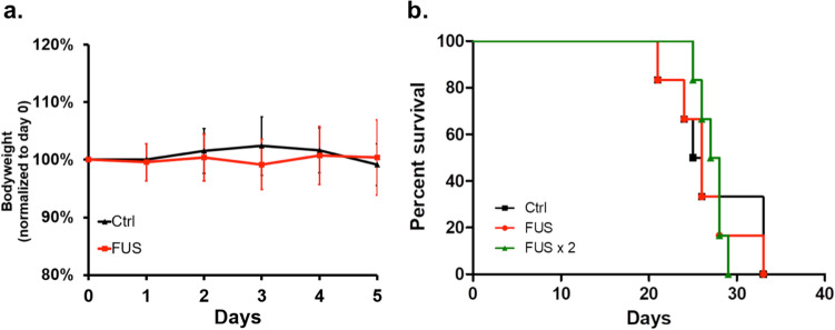
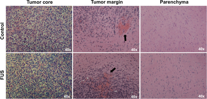
Drug delivery in diffuse intrinsic pontine glioma is significantly limited by the blood-brain barrier (BBB). Focused ultrasound (FUS), when combined with the administration of microbubbles can effectively open the BBB permitting the entry of drugs across the cerebrovasculature into the brainstem. Given that the utility of FUS in brainstem malignancies remains unknown, the purpose of our study was to determine the safety and feasibility of this technique in a murine pontine glioma model. A syngeneic orthotopic model was developed by stereotactic injection of PDGF-BPTENp53 murine glioma cells into the pons of B6 mice. A single-element, spherical-segment 1.5 MHz ultrasound transducer driven by a function generator through a power amplifier was used with concurrent intravenous microbubble injection for tumor sonication. Mice were randomly assigned to control, FUS and double-FUS groups. Pulse and respiratory rates were continuously monitored during treatment. BBB opening was confirmed with gadolinium-enhanced MRI and Evans blue. Kondziela inverted screen testing and sequential weight lifting measured motor function before and after sonication. A subset of animals were treated with etoposide following ultrasound. Mice were either sacrificed for tissue analysis or serially monitored for survival with daily weights. FUS successfully caused BBB opening while preserving normal cardiorespiratory and motor function. Furthermore, the degree of intra-tumoral hemorrhage and inflammation on H&E in control and treated mice was similar. There was also no difference in weight loss and survival between the groups (p > 0.05). Lastly, FUS increased intra-tumoral etoposide concentration by more than fivefold. FUS is a safe and feasible technique for repeated BBB opening and etoposide delivery in a preclinical pontine glioma model.
弥漫性脑桥胶质瘤中的药物递送受到血脑屏障(BBB)的显著限制。聚焦超声(FUS)与微泡联合给药时,可有效打开血脑屏障,使药物通过脑血管进入脑干。鉴于FUS在脑干恶性肿瘤中的应用效果尚不清楚,我们研究的目的是确定该技术在小鼠脑桥胶质瘤模型中的安全性和可行性。通过将PDGF - BPTENp53小鼠胶质瘤细胞立体定向注射到B6小鼠的脑桥中,建立了同基因原位模型。使用由函数发生器通过功率放大器驱动的单元素球形段1.5 MHz超声换能器,同时静脉注射微泡进行肿瘤超声处理。将小鼠随机分为对照组、FUS组和双FUS组。治疗期间持续监测脉搏和呼吸频率。通过钆增强MRI和伊文思蓝证实血脑屏障开放。在超声处理前后,通过Kondziela倒屏试验和连续举重测量运动功能。超声处理后,对一部分动物给予依托泊苷治疗。对小鼠进行处死以进行组织分析,或通过每日称重连续监测生存期。FUS成功地打开了血脑屏障,同时保留了正常的心肺和运动功能。此外,对照组和治疗组小鼠的H&E染色显示肿瘤内出血和炎症程度相似。各组之间体重减轻和生存期也无差异(p>0.05)。最后,FUS使肿瘤内依托泊苷浓度增加了五倍以上。在临床前脑桥胶质瘤模型中,FUS是一种安全可行的技术,可用于重复打开血脑屏障和递送依托泊苷。