Kim Sung-Jae, Koh Yong Gon, Kim Yong Sang
Department of Orthopaedic Surgery, Center for Stem Cell & Arthritis Research, Yonsei Sarang Hospital, 10, Hyoryeong-ro, Seocho-gu, Seoul, 06698, Republic of Korea.
BMC Musculoskelet Disord. 2021 Mar 24;22(1):301. doi: 10.1186/s12891-021-04183-y.
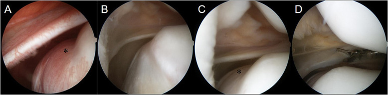
An inflamed and thickened medial patellar plica (MPP) caused by repeated mechanical irritation from trauma or overuse leads to impingement between the anterior medial femoral condyle and the medial articular facet of the patella and produces pain or clicking, which is known as MPP syndrome. In patients with MPP syndrome, cartilage damage may occur depending on the shape of the MPP and the duration of the impingement.
Preoperative magnetic resonance imaging in a 17-year-old male patient with MPP syndrome showed a hypertrophic MPP along with an abnormal notch in the articular surface of the medial femoral condyle. We considered that the impinged hypertrophic plica between the anterior medial femoral condyle and the medial articular facet of the patella resulted in cartilage damage on the articular surface of the medial femoral condyle. However, during arthroscopic surgery, we found that the cartilage of the notch, which was located beneath the MPP, was completely intact. We concluded that this abnormal notch had developed gradually in the MPP without cartilage damage.
Surgeons should be mindful that acquired plica-induced notches in the articular surface of the medial femoral condyle can present in patients with MPP syndrome.
创伤或过度使用导致的反复机械刺激引起内侧髌滑膜皱襞(MPP)发炎和增厚,进而导致股骨内侧髁前部与髌骨内侧关节面之间发生撞击,产生疼痛或弹响,这被称为MPP综合征。在MPP综合征患者中,根据MPP的形状和撞击持续时间,可能会发生软骨损伤。
一名17岁男性MPP综合征患者的术前磁共振成像显示,MPP肥厚,同时股骨内侧髁关节面有异常切迹。我们认为,股骨内侧髁前部与髌骨内侧关节面之间受撞击的肥厚滑膜皱襞导致了股骨内侧髁关节面的软骨损伤。然而,在关节镜手术中,我们发现位于MPP下方切迹处的软骨完全完整。我们得出结论,这个异常切迹是在MPP中逐渐形成的,没有软骨损伤。
外科医生应注意,MPP综合征患者可能出现后天性滑膜皱襞引起的股骨内侧髁关节面切迹。