• 文献检索
  • 文档翻译
  • 深度研究
  • 学术资讯
  • Suppr Zotero 插件Zotero 插件
  • 邀请有礼
  • 套餐&价格
  • 历史记录
应用&插件
Suppr Zotero 插件Zotero 插件浏览器插件Mac 客户端Windows 客户端微信小程序
定价
高级版会员购买积分包购买API积分包
服务
文献检索文档翻译深度研究API 文档MCP 服务
关于我们
关于 Suppr公司介绍联系我们用户协议隐私条款
关注我们

Suppr 超能文献

核心技术专利:CN118964589B侵权必究
粤ICP备2023148730 号-1Suppr @ 2026

文献检索

告别复杂PubMed语法,用中文像聊天一样搜索,搜遍4000万医学文献。AI智能推荐,让科研检索更轻松。

立即免费搜索

文件翻译

保留排版,准确专业,支持PDF/Word/PPT等文件格式,支持 12+语言互译。

免费翻译文档

深度研究

AI帮你快速写综述,25分钟生成高质量综述,智能提取关键信息,辅助科研写作。

立即免费体验

NASH 限制了免疫治疗治疗 HCC 的肿瘤监测。

NASH limits anti-tumour surveillance in immunotherapy-treated HCC.

机构信息

Division of Chronic Inflammation and Cancer, German Cancer Research Center (DKFZ), Heidelberg, Germany.

Liver Disease Research, Global Drug Discovery, Novo Nordisk A/S, Malov, Denmark.

出版信息

Nature. 2021 Apr;592(7854):450-456. doi: 10.1038/s41586-021-03362-0. Epub 2021 Mar 24.

DOI:10.1038/s41586-021-03362-0
PMID:33762733
原文链接:https://pmc.ncbi.nlm.nih.gov/articles/PMC8046670/
Abstract

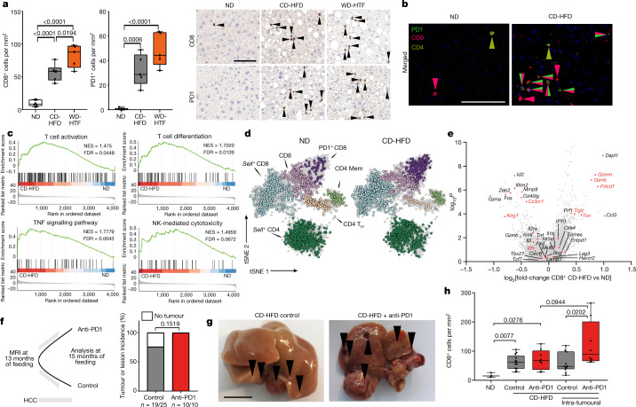
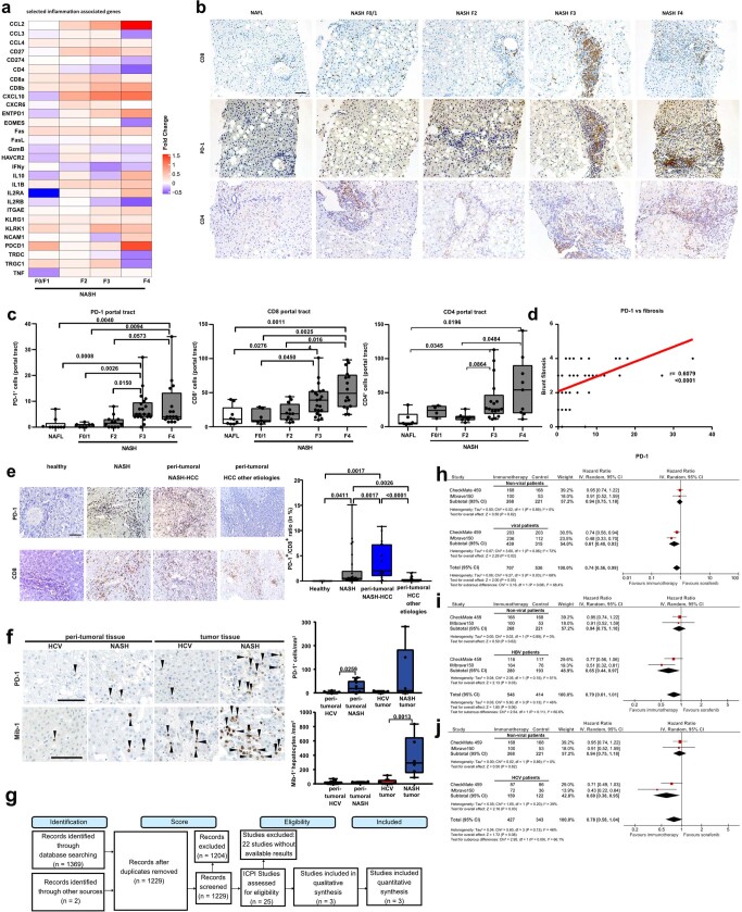
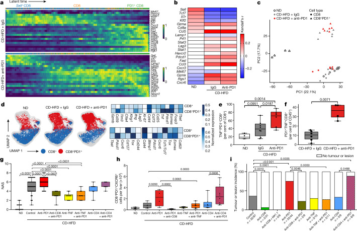
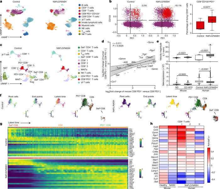
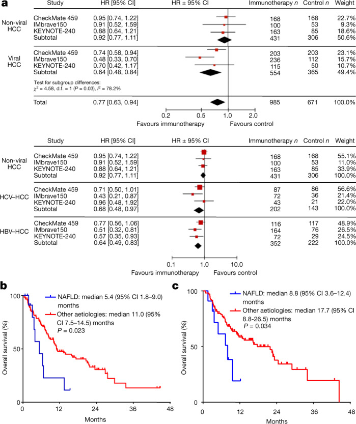
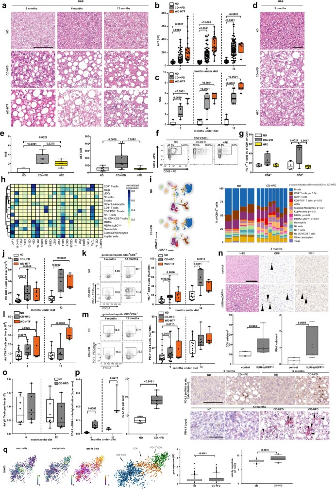
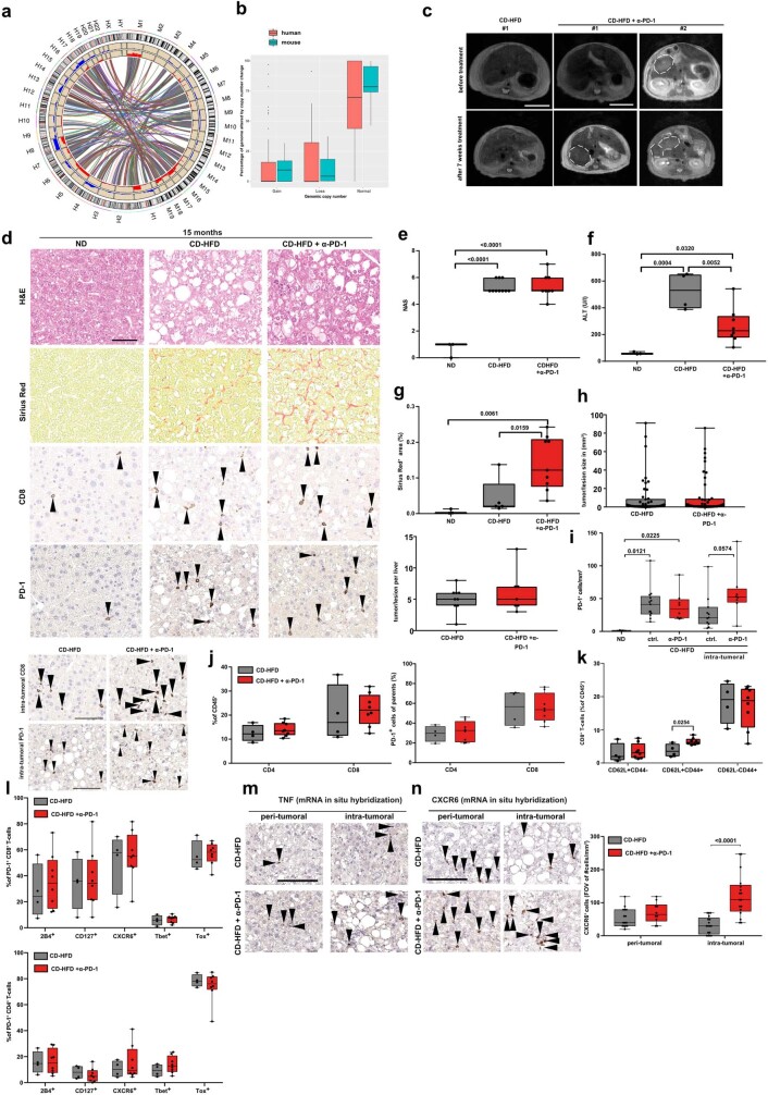
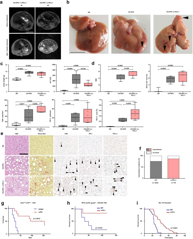
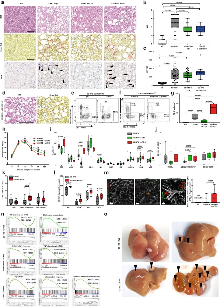
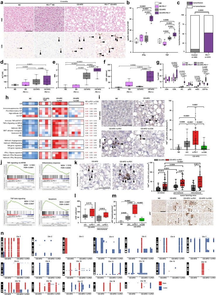
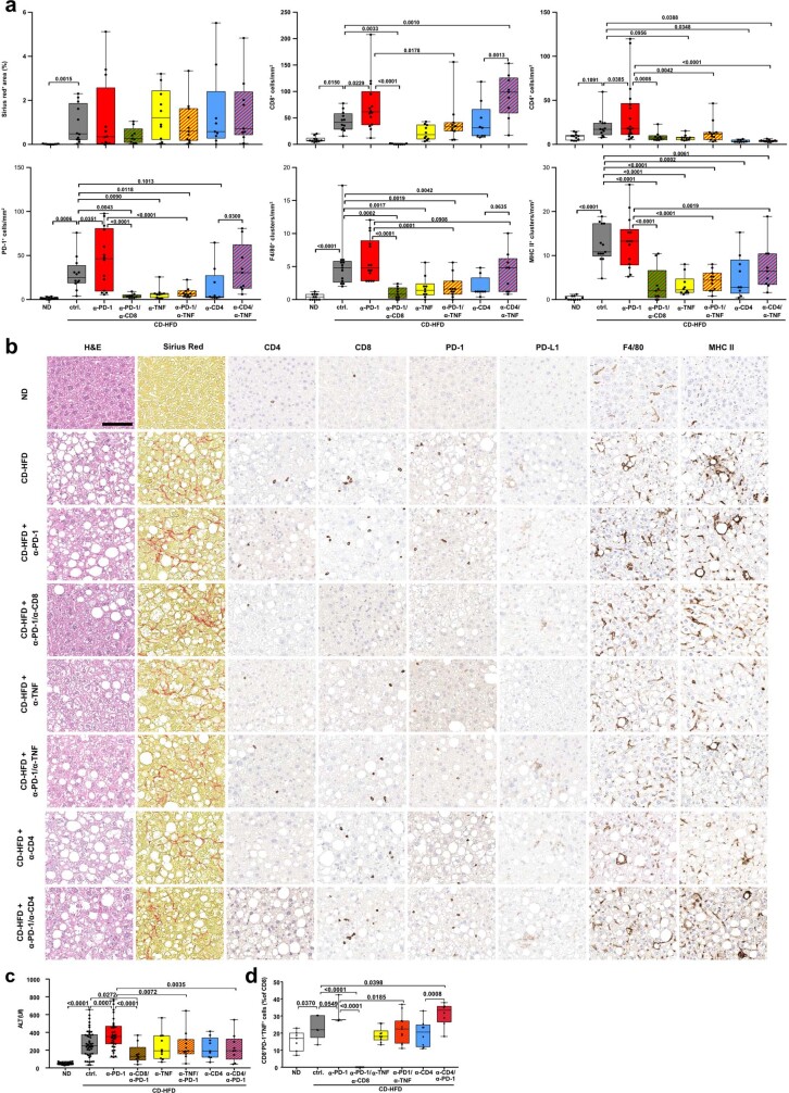
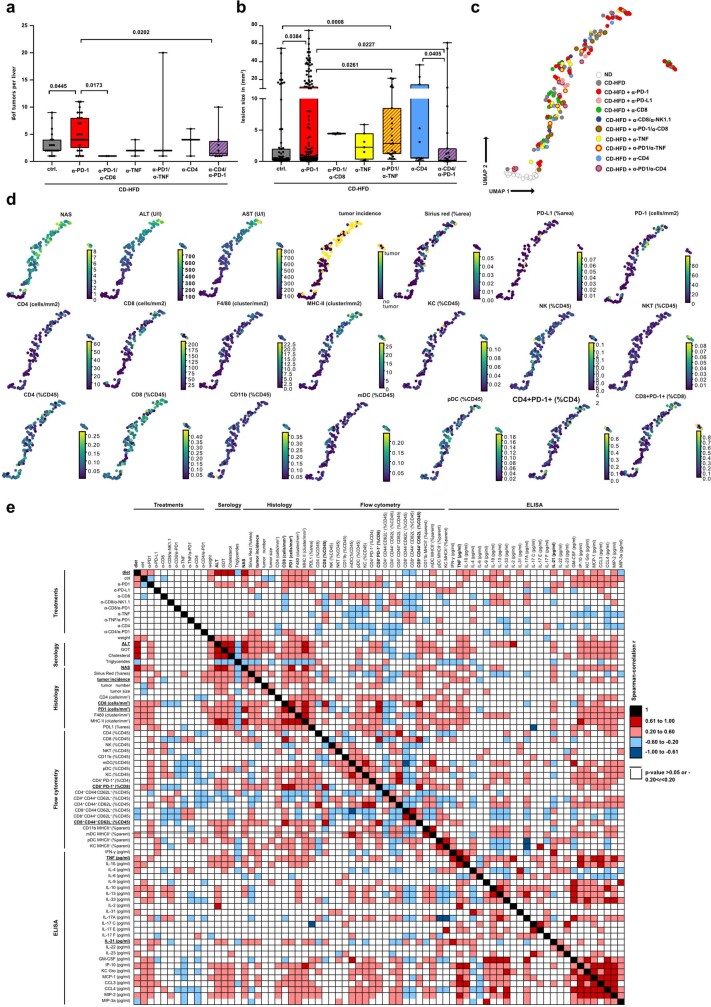
Hepatocellular carcinoma (HCC) can have viral or non-viral causes. Non-alcoholic steatohepatitis (NASH) is an important driver of HCC. Immunotherapy has been approved for treating HCC, but biomarker-based stratification of patients for optimal response to therapy is an unmet need. Here we report the progressive accumulation of exhausted, unconventionally activated CD8PD1 T cells in NASH-affected livers. In preclinical models of NASH-induced HCC, therapeutic immunotherapy targeted at programmed death-1 (PD1) expanded activated CD8PD1 T cells within tumours but did not lead to tumour regression, which indicates that tumour immune surveillance was impaired. When given prophylactically, anti-PD1 treatment led to an increase in the incidence of NASH-HCC and in the number and size of tumour nodules, which correlated with increased hepatic CD8PD1CXCR6, TOX, and TNF T cells. The increase in HCC triggered by anti-PD1 treatment was prevented by depletion of CD8 T cells or TNF neutralization, suggesting that CD8 T cells help to induce NASH-HCC, rather than invigorating or executing immune surveillance. We found similar phenotypic and functional profiles in hepatic CD8PD1 T cells from humans with NAFLD or NASH. A meta-analysis of three randomized phase III clinical trials that tested inhibitors of PDL1 (programmed death-ligand 1) or PD1 in more than 1,600 patients with advanced HCC revealed that immune therapy did not improve survival in patients with non-viral HCC. In two additional cohorts, patients with NASH-driven HCC who received anti-PD1 or anti-PDL1 treatment showed reduced overall survival compared to patients with other aetiologies. Collectively, these data show that non-viral HCC, and particularly NASH-HCC, might be less responsive to immunotherapy, probably owing to NASH-related aberrant T cell activation causing tissue damage that leads to impaired immune surveillance. Our data provide a rationale for stratification of patients with HCC according to underlying aetiology in studies of immunotherapy as a primary or adjuvant treatment.

摘要

肝细胞癌(HCC)可由病毒或非病毒因素引起。非酒精性脂肪性肝炎(NASH)是 HCC 的重要驱动因素。免疫疗法已被批准用于治疗 HCC,但基于生物标志物对患者进行分层以实现最佳治疗反应是未满足的需求。在这里,我们报告了在 NASH 影响的肝脏中耗尽的、非常规激活的 CD8PD1 T 细胞的逐渐积累。在 NASH 诱导的 HCC 的临床前模型中,针对程序性死亡受体 1(PD1)的治疗性免疫疗法在肿瘤内扩增了激活的 CD8PD1 T 细胞,但并未导致肿瘤消退,这表明肿瘤免疫监视受损。预防性给予抗 PD1 治疗会导致 NASH-HCC 的发生率增加,肿瘤结节的数量和大小增加,这与肝内 CD8PD1CXCR6、TOX 和 TNF T 细胞的增加相关。抗 PD1 治疗引发的 HCC 增加可通过耗尽 CD8 T 细胞或 TNF 中和来预防,这表明 CD8 T 细胞有助于诱导 NASH-HCC,而不是增强或执行免疫监视。我们在患有 NAFLD 或 NASH 的人类肝脏中的 CD8PD1 T 细胞中发现了类似的表型和功能特征。对超过 1600 名晚期 HCC 患者进行的三种 PD-L1(程序性死亡配体 1)或 PD1 抑制剂的随机 III 期临床试验的荟萃分析表明,免疫疗法并未改善非病毒性 HCC 患者的生存率。在另外两个队列中,接受抗 PD1 或抗 PD-L1 治疗的 NASH 驱动 HCC 患者的总生存期较其他病因患者缩短。总的来说,这些数据表明非病毒性 HCC,特别是 NASH-HCC,可能对免疫疗法的反应较差,可能是由于 NASH 相关的异常 T 细胞激活导致组织损伤,从而导致免疫监视受损。我们的数据为根据 HCC 患者的潜在病因对接受免疫治疗作为一线或辅助治疗的 HCC 患者进行分层提供了依据。

https://cdn.ncbi.nlm.nih.gov/pmc/blobs/d2d2/8046670/88204ed3fe4d/41586_2021_3362_Fig14_ESM.jpg
https://cdn.ncbi.nlm.nih.gov/pmc/blobs/d2d2/8046670/1e3304d633ee/41586_2021_3362_Fig1_HTML.jpg
https://cdn.ncbi.nlm.nih.gov/pmc/blobs/d2d2/8046670/01db7f245d67/41586_2021_3362_Fig2_HTML.jpg
https://cdn.ncbi.nlm.nih.gov/pmc/blobs/d2d2/8046670/e2623b39f4f8/41586_2021_3362_Fig3_HTML.jpg
https://cdn.ncbi.nlm.nih.gov/pmc/blobs/d2d2/8046670/91d4b4bfdc0c/41586_2021_3362_Fig4_HTML.jpg
https://cdn.ncbi.nlm.nih.gov/pmc/blobs/d2d2/8046670/3c551f314879/41586_2021_3362_Fig5_ESM.jpg
https://cdn.ncbi.nlm.nih.gov/pmc/blobs/d2d2/8046670/7df5550f2c6c/41586_2021_3362_Fig6_ESM.jpg
https://cdn.ncbi.nlm.nih.gov/pmc/blobs/d2d2/8046670/f0cdec9bb1e6/41586_2021_3362_Fig7_ESM.jpg
https://cdn.ncbi.nlm.nih.gov/pmc/blobs/d2d2/8046670/6fc6451ea1af/41586_2021_3362_Fig8_ESM.jpg
https://cdn.ncbi.nlm.nih.gov/pmc/blobs/d2d2/8046670/c3b916498528/41586_2021_3362_Fig9_ESM.jpg
https://cdn.ncbi.nlm.nih.gov/pmc/blobs/d2d2/8046670/ac47df99fdf0/41586_2021_3362_Fig10_ESM.jpg
https://cdn.ncbi.nlm.nih.gov/pmc/blobs/d2d2/8046670/5a4461bdcc6b/41586_2021_3362_Fig11_ESM.jpg
https://cdn.ncbi.nlm.nih.gov/pmc/blobs/d2d2/8046670/f6a561dc43bd/41586_2021_3362_Fig12_ESM.jpg
https://cdn.ncbi.nlm.nih.gov/pmc/blobs/d2d2/8046670/4a413228935a/41586_2021_3362_Fig13_ESM.jpg
https://cdn.ncbi.nlm.nih.gov/pmc/blobs/d2d2/8046670/88204ed3fe4d/41586_2021_3362_Fig14_ESM.jpg

相似文献

1
NASH limits anti-tumour surveillance in immunotherapy-treated HCC.NASH 限制了免疫治疗治疗 HCC 的肿瘤监测。
Nature. 2021 Apr;592(7854):450-456. doi: 10.1038/s41586-021-03362-0. Epub 2021 Mar 24.
2
The Role of CD4T Cells in Nonalcoholic Steatohepatitis and Hepatocellular Carcinoma.CD4T 细胞在非酒精性脂肪性肝炎和肝细胞癌中的作用。
Int J Mol Sci. 2024 Jun 23;25(13):6895. doi: 10.3390/ijms25136895.
3
TOX promotes the exhaustion of antitumor CD8 T cells by preventing PD1 degradation in hepatocellular carcinoma.TOX 通过阻止 PD1 降解促进肝癌中抗肿瘤 CD8 T 细胞耗竭。
J Hepatol. 2019 Oct;71(4):731-741. doi: 10.1016/j.jhep.2019.05.015. Epub 2019 Jun 5.
4
Anti-4-1BB×PDL1 Bispecific Antibody Reinvigorates Tumor-Specific Exhausted CD8+ T Cells and Enhances the Efficacy of Anti-PD1 Blockade.抗 4-1BB×PDL1 双特异性抗体使肿瘤特异性耗竭 CD8+ T 细胞重新焕发活力,并增强抗 PD1 阻断的疗效。
Clin Cancer Res. 2024 Sep 13;30(18):4155-4166. doi: 10.1158/1078-0432.CCR-23-2864.
5
Association Between Expression Level of PD1 by Tumor-Infiltrating CD8 T Cells and Features of Hepatocellular Carcinoma.肿瘤浸润 CD8 T 细胞中 PD1 的表达水平与肝细胞癌特征的关系。
Gastroenterology. 2018 Dec;155(6):1936-1950.e17. doi: 10.1053/j.gastro.2018.08.030. Epub 2018 Aug 24.
6
Anti-CD122 antibody restores specific CD8 T cell response in nonalcoholic steatohepatitis and prevents hepatocellular carcinoma growth.抗 CD122 抗体可恢复非酒精性脂肪性肝炎中特异性 CD8 T 细胞应答,并防止肝细胞癌生长。
Oncoimmunology. 2023 Mar 2;12(1):2184991. doi: 10.1080/2162402X.2023.2184991. eCollection 2023.
7
PD1 CD8 T cells correlate with exhausted signature and poor clinical outcome in hepatocellular carcinoma.PD1 CD8 T 细胞与肝癌衰竭特征和不良临床结局相关。
J Immunother Cancer. 2019 Nov 29;7(1):331. doi: 10.1186/s40425-019-0814-7.
8
Lysine-specific demethylase 1 deletion reshapes tumour microenvironment to overcome acquired resistance to anti-programmed death 1 therapy in liver cancer.赖氨酸特异性去甲基化酶1缺失重塑肿瘤微环境以克服肝癌中对程序性死亡1疗法的获得性耐药。
Clin Transl Med. 2025 May;15(5):e70335. doi: 10.1002/ctm2.70335.
9
Disruption of SIRT7 Increases the Efficacy of Checkpoint Inhibitor via MEF2D Regulation of Programmed Cell Death 1 Ligand 1 in Hepatocellular Carcinoma Cells.SIRT7 缺失通过 MEF2D 调控程序性细胞死亡配体 1 增加肝癌细胞中检查点抑制剂的疗效。
Gastroenterology. 2020 Feb;158(3):664-678.e24. doi: 10.1053/j.gastro.2019.10.025. Epub 2019 Oct 31.
10
TIGIT and PD1 Co-blockade Restores ex vivo Functions of Human Tumor-Infiltrating CD8 T Cells in Hepatocellular Carcinoma.TIGIT 和 PD1 联合阻断恢复肝癌患者肿瘤浸润 CD8 T 细胞的体外功能。
Cell Mol Gastroenterol Hepatol. 2021;12(2):443-464. doi: 10.1016/j.jcmgh.2021.03.003. Epub 2021 Mar 27.

引用本文的文献

1
Real-World Outcomes of Atezolizumab-Bevacizumab in Hepatocellular Carcinoma: The Prospective French CHIEF Cohort.阿替利珠单抗联合贝伐单抗治疗肝细胞癌的真实世界疗效:法国前瞻性CHIEF队列研究
Liver Int. 2025 Oct;45(10):e70337. doi: 10.1111/liv.70337.
2
Global, regional, and national burden of NASH related liver cancer in adults aged 45 and above: an analysis from the GBD 2021 and forecast to 2050.45岁及以上成年人中与非酒精性脂肪性肝炎相关的肝癌的全球、区域和国家负担:来自全球疾病负担研究(GBD)2021的分析及到2050年的预测
Front Nutr. 2025 Aug 19;12:1651357. doi: 10.3389/fnut.2025.1651357. eCollection 2025.
3
Single-cell atlas of human liver and blood immune cells across fatty liver disease stages reveals distinct signatures linked to liver dysfunction and fibrogenesis.

本文引用的文献

1
Hepatocellular carcinoma.肝细胞癌。
Nat Rev Dis Primers. 2021 Jan 21;7(1):6. doi: 10.1038/s41572-020-00240-3.
2
Transcriptomic profiling across the nonalcoholic fatty liver disease spectrum reveals gene signatures for steatohepatitis and fibrosis.非酒精性脂肪性肝病谱中的转录组特征分析揭示了肝脂肪变性和纤维化的基因特征。
Sci Transl Med. 2020 Dec 2;12(572). doi: 10.1126/scitranslmed.aba4448.
3
The European NAFLD Registry: A real-world longitudinal cohort study of nonalcoholic fatty liver disease.《欧洲非酒精性脂肪性肝病注册研究:一项真实世界的非酒精性脂肪性肝病纵向队列研究》。
跨越脂肪肝疾病各阶段的人类肝脏和血液免疫细胞单细胞图谱揭示了与肝功能障碍和纤维化相关的独特特征。
Nat Immunol. 2025 Sep;26(9):1596-1611. doi: 10.1038/s41590-025-02255-y. Epub 2025 Aug 29.
4
Steatohepatitis alters lymphocytes cytotoxicity and localization, accelerating colorectal liver metastases.脂肪性肝炎会改变淋巴细胞的细胞毒性和定位,加速结直肠癌肝转移。
Neoplasia. 2025 Aug 28;69:101222. doi: 10.1016/j.neo.2025.101222.
5
Immunological Landscape and Molecular Therapeutic Targets of the Tumor Microenvironment in Hepatocellular Carcinoma.肝细胞癌肿瘤微环境的免疫格局与分子治疗靶点
Int J Mol Sci. 2025 Aug 13;26(16):7836. doi: 10.3390/ijms26167836.
6
Radiomics-based prediction of HCC response to atezolizumab/bevacizumab.基于影像组学预测肝细胞癌对阿替利珠单抗/贝伐单抗的反应
Oncol Lett. 2025 Aug 14;30(4):484. doi: 10.3892/ol.2025.15229. eCollection 2025 Oct.
7
Current knowledge about immunotherapy response after liver transplantation of patients with liver cancer.关于肝癌患者肝移植后免疫治疗反应的当前知识。
J Liver Transpl. 2025 Aug;19. doi: 10.1016/j.liver.2025.100285. Epub 2025 Jun 6.
8
Serum fibrosis marker M2BPGi-based novel score predicts survival of unresectable HCC undergoing immunotherapy.基于血清纤维化标志物M2BPGi的新型评分可预测接受免疫治疗的不可切除肝癌患者的生存率。
JHEP Rep. 2025 Jun 26;7(9):101491. doi: 10.1016/j.jhepr.2025.101491. eCollection 2025 Sep.
9
Reevaluating the Role of Demographic and Etiological Factors in Immune Checkpoint Inhibitor Therapy for Advanced Hepatocellular Carcinoma.重新评估人口统计学和病因学因素在晚期肝细胞癌免疫检查点抑制剂治疗中的作用
Liver Cancer. 2025 Mar 3;14(4):508-510. doi: 10.1159/000544964. eCollection 2025 Aug.
10
Treatment Duration of Adjuvant Immune Checkpoint Inhibitors in Hepatocellular Carcinoma Patients at High Risk of Recurrence after Resection: A Prospective, Multicentric Cohort Study.肝细胞癌切除术后复发高危患者辅助免疫检查点抑制剂的治疗持续时间:一项前瞻性、多中心队列研究
Liver Cancer. 2024 Dec 4;14(4):378-390. doi: 10.1159/000542954. eCollection 2025 Aug.
Contemp Clin Trials. 2020 Nov;98:106175. doi: 10.1016/j.cct.2020.106175. Epub 2020 Oct 9.
4
Steatohepatitis Impairs T-cell-Directed Immunotherapies Against Liver Tumors in Mice.脂肪性肝炎损害小鼠针对肝肿瘤的T细胞导向免疫疗法。
Gastroenterology. 2021 Jan;160(1):331-345.e6. doi: 10.1053/j.gastro.2020.09.031. Epub 2020 Oct 1.
5
Post-registration experience of nivolumab in advanced hepatocellular carcinoma: an international study.纳武利尤单抗在晚期肝细胞癌中的注册后经验:一项国际研究。
J Immunother Cancer. 2020 Aug;8(2). doi: 10.1136/jitc-2020-001033.
6
Hyperprogressive disease during PD-1 blockade in patients with advanced hepatocellular carcinoma.抗 PD-1 治疗期间晚期肝细胞癌患者的超进展性疾病。
J Hepatol. 2021 Feb;74(2):350-359. doi: 10.1016/j.jhep.2020.08.010. Epub 2020 Aug 15.
7
Generalizing RNA velocity to transient cell states through dynamical modeling.通过动态建模将 RNA 速度推广到瞬时细胞状态。
Nat Biotechnol. 2020 Dec;38(12):1408-1414. doi: 10.1038/s41587-020-0591-3. Epub 2020 Aug 3.
8
Immunotherapy for advanced hepatocellular carcinoma: a focus on special subgroups.免疫疗法治疗晚期肝细胞癌:关注特殊亚组。
Gut. 2021 Jan;70(1):204-214. doi: 10.1136/gutjnl-2020-321702. Epub 2020 Aug 3.
9
Association of inflammatory biomarkers with clinical outcomes in nivolumab-treated patients with advanced hepatocellular carcinoma.纳武利尤单抗治疗晚期肝细胞癌患者的炎症生物标志物与临床结局的关系。
J Hepatol. 2020 Dec;73(6):1460-1469. doi: 10.1016/j.jhep.2020.07.026. Epub 2020 Jul 22.
10
Atezolizumab plus Bevacizumab in Unresectable Hepatocellular Carcinoma.阿替利珠单抗联合贝伐珠单抗治疗不可切除肝细胞癌。
N Engl J Med. 2020 May 14;382(20):1894-1905. doi: 10.1056/NEJMoa1915745.