Chen Amei, Huang Jun-Xiang, Liao Yuting, Liu Zaosong, Chen Dandan, Yang Chongzhe, Yang Rui-Meng, Wei Xinhua
Department of Radiology, Guangzhou First Hospital, The Second Affiliated Hospital, South China University of Technology, No. 1 Panfu Road, Guangzhou 510180, China (A.C., Z.L., D.C., R.Y., X.W.); Department of Anesthesiology, Guangzhou Women and Children's Medical Center, Guangzhou, Guangdong, China (J.H.); GE Healthcare, Guangzhou, China (Y.L.); and Department of Geriatrics, Guangzhou First People's Hospital, The Second Affiliated Hospital, South China University of Technology, Guangzhou, China (C.Y.).
Radiol Cardiothorac Imaging. 2020 Apr 6;2(2):e200117. doi: 10.1148/ryct.2020200117. eCollection 2020 Apr.
To characterize and compare the initial clinical and imaging features of coronavirus disease 2019 (COVID-19) in pediatric and adult patients undergoing chest CT.
A total of 61 patients, consisting of 47 adults (aged 18 years or older) and 14 pediatric patients (aged younger than 18 years) with laboratory-confirmed COVID-19 confirmed by real-time reverse-transcription polymerase chain reaction between January 25 and February 15, 2020, were enrolled in this study. All patients underwent chest CT within 3 days after the initial reverse transcription polymerase chain reaction test. The clinical presentation, serum markers, and CT findings were assessed and compared between the adult and pediatric patients.
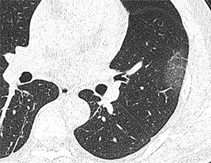
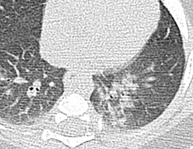
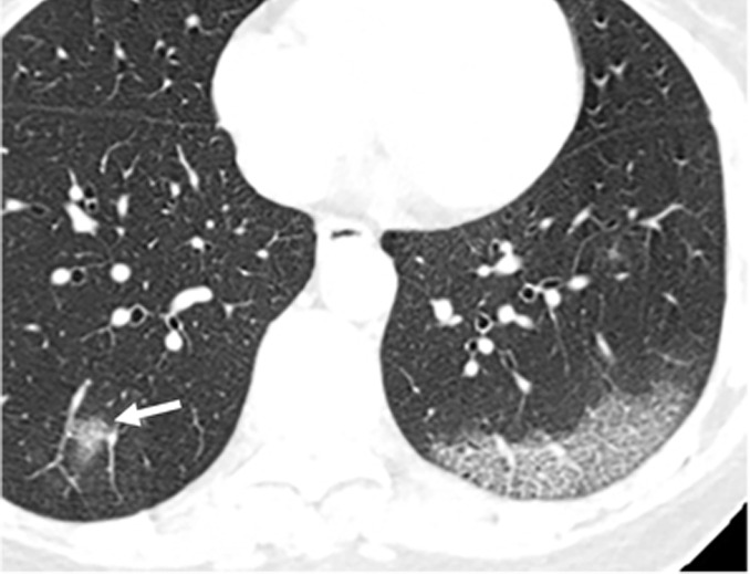
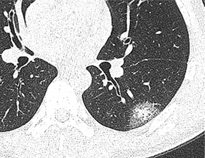
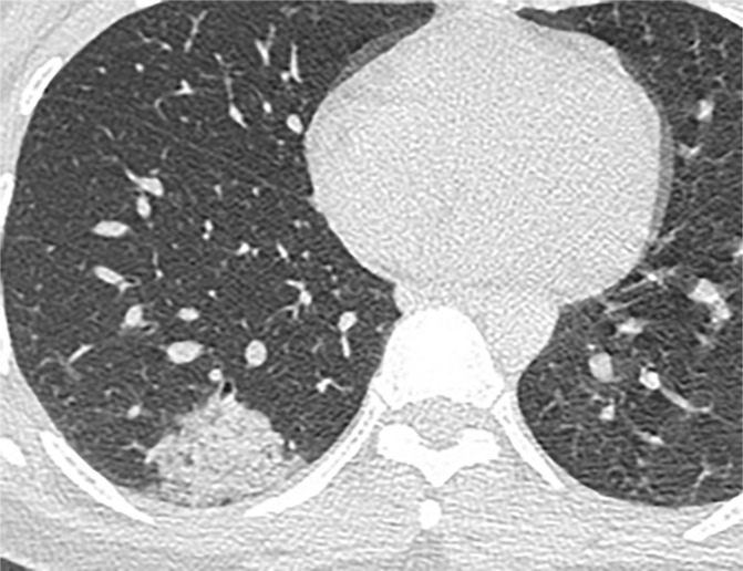
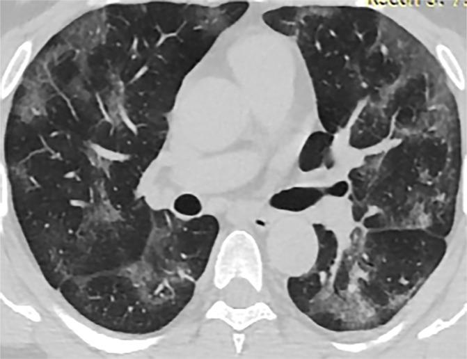
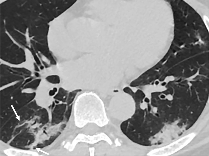
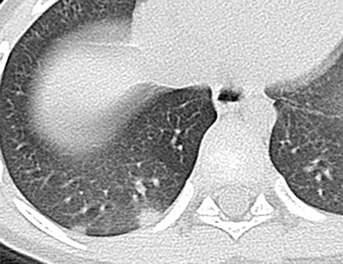
Fever was less common in pediatric patients than in adults (six of 14, 42.9% vs 39 of 47, 83%; = .008). Leukopenia or normal, lymphopenia or normal, and increased or normal C-reactive protein level were common in both groups with no difference ( > .05). Compared with the adults, pediatric patients had a lower rate of positive CT findings and a milder clinical grade ( = .004 and = .001, respectively). At chest CT, the number of pulmonary lobes involved was found to be reduced in pediatric patients when compared with adults ( = .012). Subpleural distribution of lung opacities was a dominant feature in both groups, whereas bronchial distribution was more common in the pediatric group ( = .048). Among the CT features in adults, ground-glass opacities (GGOs) were the most common finding (24 of 43, 53.5%), followed by GGO with consolidation (14 of 43, 27.9%). In pediatric patients, GGOs accounted for 42.9% (three of seven), bronchial wall thickening occurred in 28.6% (two of seven), and GGOs with consolidations and nodular opacities occurred in 14.3% (one of seven). However, these CT features did not differ in the two groups, except for bronchial wall thickening, which was more commonly found in pediatric patients ( = .048). In addition, the semiquantitative scores of lung involvement were higher in adults than in pediatric patients (8.89 ± 4.54 vs 1.86 ± 2.41; < .001).
Compared with adults, pediatric patients with COVID-19 showed distinctive clinical and CT features. Pediatric patients tend to have milder clinical symptoms, fewer positive results at CT, and less extensive involvement at imaging. Bronchial wall thickening was relatively more frequent on CT images from pediatric patients with COVID-19 in comparison with adults.© RSNA, 2020.
对接受胸部CT检查的儿童和成年2019冠状病毒病(COVID-19)患者的初始临床和影像特征进行描述及比较。
本研究纳入了2020年1月25日至2月15日期间经实时逆转录聚合酶链反应实验室确诊为COVID-19的61例患者,其中包括47例成年人(年龄18岁及以上)和14例儿童患者(年龄小于18岁)。所有患者在首次逆转录聚合酶链反应检测后的3天内接受了胸部CT检查。对成年和儿童患者的临床表现、血清标志物及CT表现进行了评估和比较。
发热在儿童患者中比在成人中少见(14例中的6例,42.9% 对比47例中的39例,83%;P = .008)。两组患者白细胞减少或正常、淋巴细胞减少或正常以及C反应蛋白水平升高或正常的情况均常见,且无差异(P > .05)。与成年人相比,儿童患者CT阳性表现率较低,临床分级较轻(分别为P = .004和P = .001)。胸部CT检查时,发现儿童患者受累肺叶数量比成年人减少(P = .012)。两组患者肺实变影均以胸膜下分布为主,而支气管周围分布在儿童组中更常见(P = .048)。在成年人的CT表现中,磨玻璃影(GGO)最为常见(43例中的24例,53.5%),其次是GGO合并实变(43例中的14例,27.9%)。在儿童患者中,GGO占42.9%(7例中的3例);支气管壁增厚占28.6%(7例中的2例);GGO合并实变及结节状实变影占14.3%(7例中的1例)。然而,除支气管壁增厚在儿童患者中更常见外(P = .048),这些CT表现两组间无差异。此外,成年人肺部受累的半定量评分高于儿童患者(8.89 ± 4.54对比1.86 ± 2.41;P < .001)。
与成年人相比,COVID-19儿童患者表现出独特的临床和CT特征。儿童患者往往临床症状较轻,CT阳性结果较少,影像上受累范围较小。与成年人相比,COVID-19儿童患者的CT图像上支气管壁增厚相对更常见。©RSNA,2020年