From the Departments of Radiology (F. Song, N.S., F. Shan, Z.Z., J.S., Y.S.) and Infectious Disease (H.L., Y.L.), Shanghai Public Health Clinical Center, No. 2501 Caolang Road, Jinshan District, Shanghai 201508, China; Department of the Principal's Office, Fudan University, Shanghai, China (Z.Z.); Cancer Center, University of Michigan, Ann Arbor, Mich (Y.J.); and Shanghai Key Laboratory of Molecular Imaging, Shanghai, China (Y.S.).
Radiology. 2020 Apr;295(1):210-217. doi: 10.1148/radiol.2020200274. Epub 2020 Feb 6.
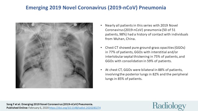
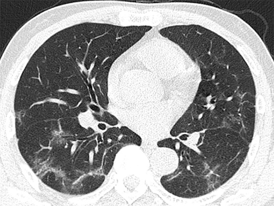
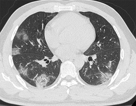
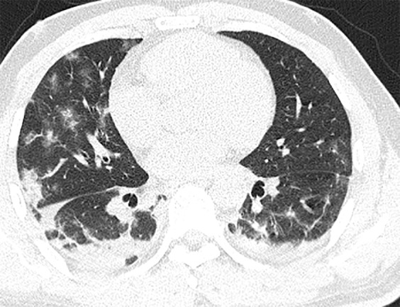
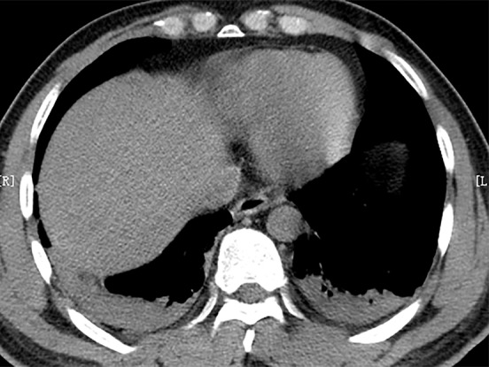
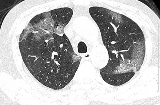
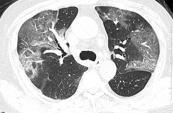
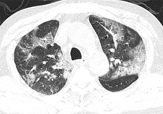
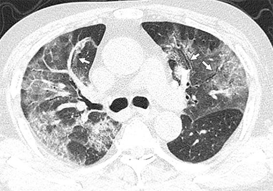
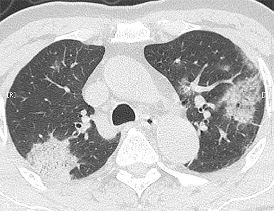
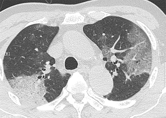
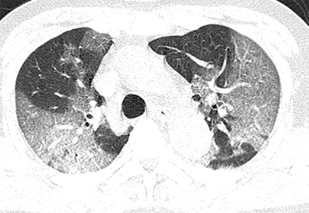
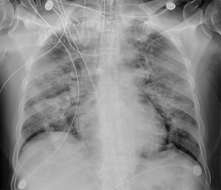
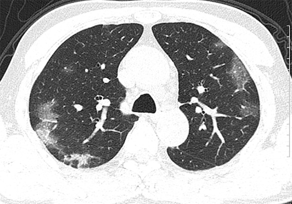
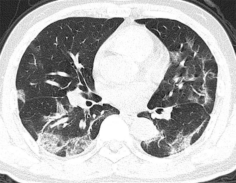
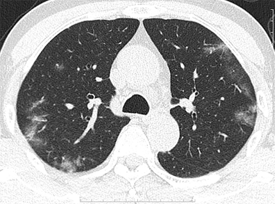
BackgroundThe chest CT findings of patients with 2019 Novel Coronavirus (2019-nCoV) pneumonia have not previously been described in detail.PurposeTo investigate the clinical, laboratory, and imaging findings of emerging 2019-nCoV pneumonia in humans.Materials and MethodsFifty-one patients (25 men and 26 women; age range 16-76 years) with laboratory-confirmed 2019-nCoV infection by using real-time reverse transcription polymerase chain reaction underwent thin-section CT. The imaging findings, clinical data, and laboratory data were evaluated.ResultsFifty of 51 patients (98%) had a history of contact with individuals from the endemic center in Wuhan, China. Fever (49 of 51, 96%) and cough (24 of 51, 47%) were the most common symptoms. Most patients had a normal white blood cell count (37 of 51, 73%), neutrophil count (44 of 51, 86%), and either normal (17 of 51, 35%) or reduced (33 of 51, 65%) lymphocyte count. CT images showed pure ground-glass opacity (GGO) in 39 of 51 (77%) patients and GGO with reticular and/or interlobular septal thickening in 38 of 51 (75%) patients. GGO with consolidation was present in 30 of 51 (59%) patients, and pure consolidation was present in 28 of 51 (55%) patients. Forty-four of 51 (86%) patients had bilateral lung involvement, while 41 of 51 (80%) involved the posterior part of the lungs and 44 of 51 (86%) were peripheral. There were more consolidated lung lesions in patients 5 days or more from disease onset to CT scan versus 4 days or fewer (431 of 712 lesions vs 129 of 612 lesions; < .001). Patients older than 50 years had more consolidated lung lesions than did those aged 50 years or younger (212 of 470 vs 198 of 854; < .001). Follow-up CT in 13 patients showed improvement in seven (54%) patients and progression in four (31%) patients.ConclusionPatients with fever and/or cough and with conspicuous ground-glass opacity lesions in the peripheral and posterior lungs on CT images, combined with normal or decreased white blood cells and a history of epidemic exposure, are highly suspected of having 2019 Novel Coronavirus (2019-nCoV) pneumonia.© RSNA, 2020.
背景 2019 年新型冠状病毒(2019-nCoV)肺炎患者的胸部 CT 表现以前尚未详细描述。
目的 探讨人类新发 2019-nCoV 肺炎的临床、实验室和影像学表现。
材料与方法 经实时逆转录聚合酶链反应检测证实有 2019-nCoV 感染的 51 例患者(25 例男性,26 例女性;年龄 16~76 岁)均接受了薄层 CT 检查。评估了影像学表现、临床数据和实验室数据。
结果 51 例患者中有 50 例(98%)有接触中国武汉流行地区人员的病史。发热(51 例,96%)和咳嗽(24 例,47%)是最常见的症状。大多数患者的白细胞计数正常(37 例,73%)、中性粒细胞计数正常(44 例,86%)或淋巴细胞计数减少(17 例,35%)或正常(33 例,65%)。CT 图像显示 51 例患者中有 39 例(77%)单纯磨玻璃样混浊(GGO),38 例(75%)GGO 伴有网状和/或小叶间隔增厚。30 例(59%)患者有 GGO 合并实变,28 例(55%)患者有单纯实变。44 例(86%)患者有双侧肺部受累,41 例(80%)累及肺部后部,44 例(86%)累及外周。与发病后 4 天或更短时间进行 CT 扫描相比,发病后 5 天或更长时间进行 CT 扫描的患者有更多的实变肺病变(712 个病变中有 431 个 vs 612 个病变中有 129 个;<.001)。50 岁以上患者的实变肺病变多于 50 岁以下患者(470 个病变中有 212 个 vs 854 个病变中有 198 个;<.001)。13 例患者的随访 CT 显示 7 例(54%)患者病情改善,4 例(31%)患者病情进展。
结论 对于有发热和/或咳嗽且 CT 图像显示外周和后部肺部有明显磨玻璃样混浊病变、白细胞正常或减少且有流行地区接触史的患者,高度怀疑患有 2019 年新型冠状病毒(2019-nCoV)肺炎。© RSNA,2020。