Department of Ophthalmology, New Zealand National Eye Centre, Faculty of Medical and Health Sciences, University of Auckland, Auckland, New Zealand.
Auckland Bioengineering Institute, University of Auckland, Auckland, New Zealand.
J Morphol. 2021 Jun;282(6):874-886. doi: 10.1002/jmor.21354. Epub 2021 May 2.
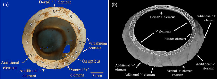
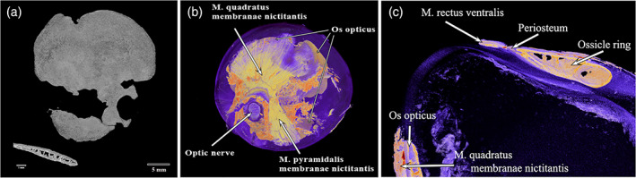
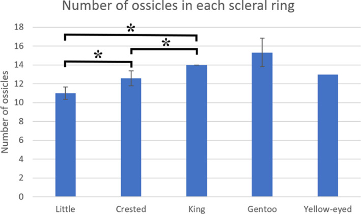
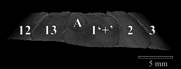
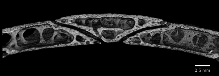
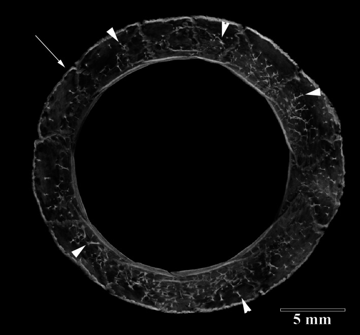
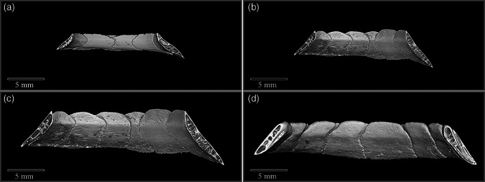
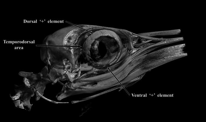
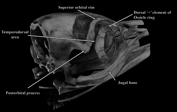
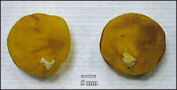
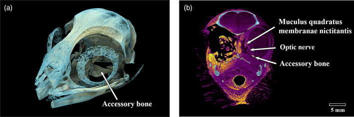
Scleral ossicles and other bony elements are present in the eyes of many vertebrates, including birds. In this study, the skeletal elements present in the penguin eye and orbit were imaged using macro photographs and micro-computed tomography (micro-CT), to help elucidate their function and significance. A total of 36 scleral rings and three whole skulls were imaged. King (Aptenodytes patagonicus), Fiordland crested (Eudyptes pachyrhynchus), Snares crested (Eudyptes robustus), royal (Eudyptes schlegeli) and yellow-eyed (Megadyptes antipodes) penguins had between 12 and 14 elements in their scleral ring while the gentoo (Pygoscelis papua) had 14 and 17; little penguins (Eudyptula sp.) consistently had between 10 and 12 elements. All had at least two elements that overlapped, usually totally, each neighbour, and two that were overlapped by each neighbour. The interior structure of all ossicles revealed a lattice-like arrangement of struts typical of cancellous bone, the whole being surrounded by thick cortical bone. The scleral ring of a 10 week gentoo chick was not completely ossified but rather had multiple small holes within it on micro-CT. A large os opticus was present in one king penguin but in another bird of the same age and gender there was no such bone. Much smaller accessory bones were found in the posterior pole of one Snares crested and one little penguin. We conclude that the penguin scleral ring not only maintains the shape of the eye but also provides protection and a site of insertion for rectus muscles. However, the extreme variability in the os opticus suggests that it is not essential to normal function.
巩膜骨小骨和其他骨骼元素存在于许多脊椎动物的眼睛中,包括鸟类。在这项研究中,使用宏观照片和微计算机断层扫描(micro-CT)对企鹅眼睛和眼眶中的骨骼元素进行成像,以帮助阐明其功能和意义。共拍摄了 36 个巩膜环和 3 个完整头骨。国王企鹅(Aptenodytes patagonicus)、菲尔德克赖斯特冠企鹅(Eudyptes pachyrhynchus)、史奈斯冠企鹅(Eudyptes robustus)、皇家企鹅(Eudyptes schlegeli)和黄眼企鹅(Megadyptes antipodes)的巩膜环中有 12 到 14 个元素,而巴布亚企鹅(Pygoscelis papua)则有 14 到 17 个;小企鹅(Eudyptula sp.)的巩膜环始终有 10 到 12 个元素。所有的都至少有两个元素相互重叠,通常是完全重叠,每个相邻的都有两个重叠。所有骨小骨的内部结构都显示出典型的海绵状骨的格子状排列,整个骨被厚厚的皮质骨包围。一只 10 周大的巴布亚企鹅的巩膜环尚未完全骨化,而是在 micro-CT 上有多个小孔。一只国王企鹅有一个大的视神经,但在同一年龄和性别的另一只鸟中没有这样的骨头。在一只史奈斯冠企鹅和一只小企鹅的后极发现了更小的副骨。我们得出结论,企鹅的巩膜环不仅维持眼睛的形状,还提供保护和直肌的插入点。然而,视神经的极端可变性表明它对正常功能不是必需的。