Department of Biology, Georgetown University, Washington, D.C. 20057, USA;
Department of Optoelectronics and Materials Engineering, Chung Hua University, Hsinchu 30012, Taiwan.
Cold Spring Harb Protoc. 2022 Jan 4;2022(1):pdb.prot106781. doi: 10.1101/pdb.prot106781.
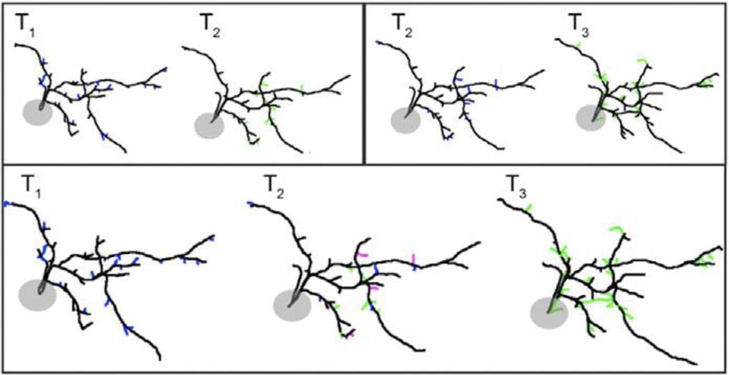
In vivo time-lapse imaging of complete dendritic arbor structures in tectal neurons of tadpoles has served as a powerful in vivo model to study activity-dependent structural plasticity in the central nervous system during early development. In addition to quantitative analysis of gross arbor structure, dynamic analysis of the four-dimensional data offers particularly valuable insights into the structural changes occurring in subcellular domains over experience/development-driven structural plasticity events. Such analysis allows not only quantifiable characterization of branch additions and retractions with high temporal resolution but also identification of the loci of action. This allows for a better understanding of the spatiotemporal association of structural changes to functional relevance. Here we describe a protocol for in vivo time-lapse imaging of complete dendritic arbors from individual neurons in the brains of anesthetized tadpoles with two-photon microscopy and data analysis of the time series of 3D dendritic arbors. For data analysis, we focus on dynamic analysis of reconstructed neuronal filaments using a customized open source computer program we developed (4D SPA), which allows aligning and matching of 3D neuronal structures across different time points with greatly improved speed and reliability. File converters are provided to convert reconstructed filament files from commercial reconstruction software to be used in 4D SPA. The program and user manual are publicly accessible and operate through a graphical user interface on both Windows and Mac OSX.
在活体延时成像中对完整的树突分支结构进行研究,为研究中枢神经系统在早期发育过程中的活性依赖的结构性可塑性提供了有力的活体模型。除了对总树突结构的定量分析外,对四维数据的动态分析特别有助于了解在经历/发育驱动的结构性可塑性事件中发生的亚细胞域的结构变化。这种分析不仅允许以高时间分辨率对分支的添加和缩回进行可量化的描述,还可以识别作用部位。这有助于更好地理解结构变化与功能相关性的时空关联。在这里,我们描述了一种使用双光子显微镜对麻醉的蝌蚪大脑中的单个神经元的完整树突分支进行在体延时成像的方案,并对三维树突分支的时间序列进行数据分析。对于数据分析,我们专注于使用我们开发的定制的开源计算机程序(4D SPA)对重建的神经元纤维进行动态分析,该程序允许以大大提高的速度和可靠性在不同时间点对齐和匹配三维神经元结构。还提供了文件转换器,用于将商业重建软件中的重建纤维文件转换为可用于 4D SPA 的格式。该程序和用户手册可公开获取,并通过图形用户界面在 Windows 和 Mac OSX 上运行。