Department of Immunology and Inflammation, Centre for Inflammatory Disease, Imperial College London, London, UK.
MRC London Institute of Medical Sciences, Imperial College London, London, UK.
Nat Commun. 2021 Mar 31;12(1):1980. doi: 10.1038/s41467-021-22312-y.
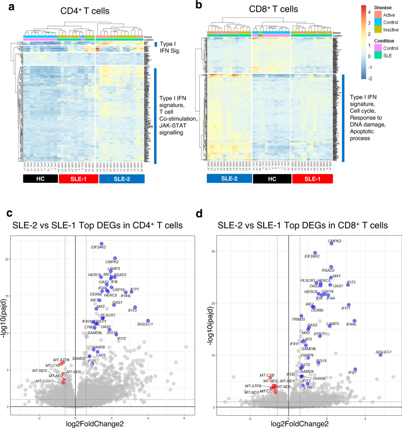
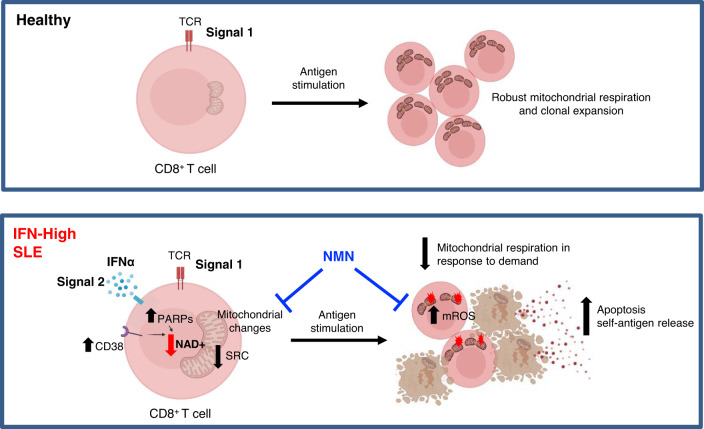
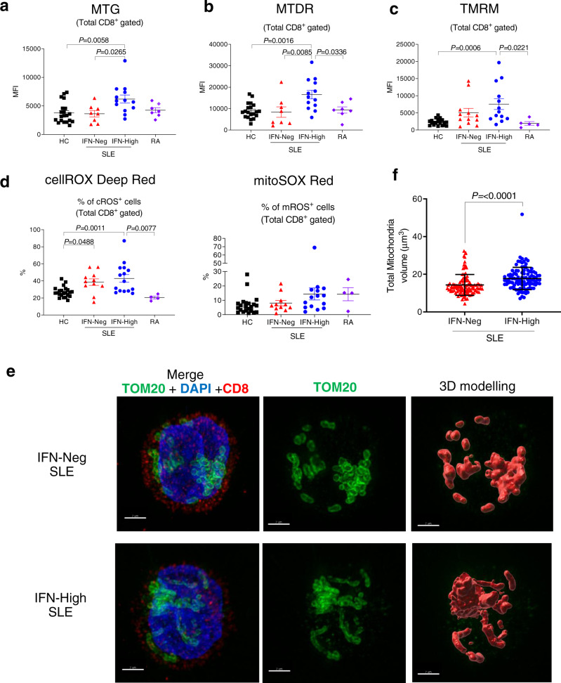
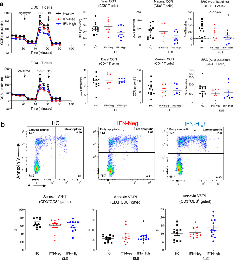
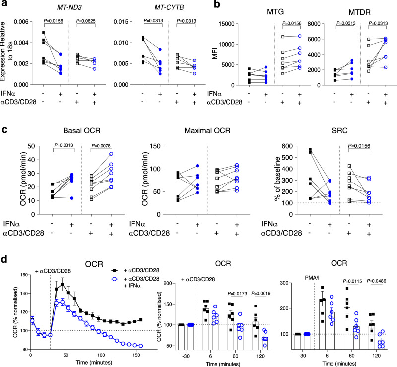
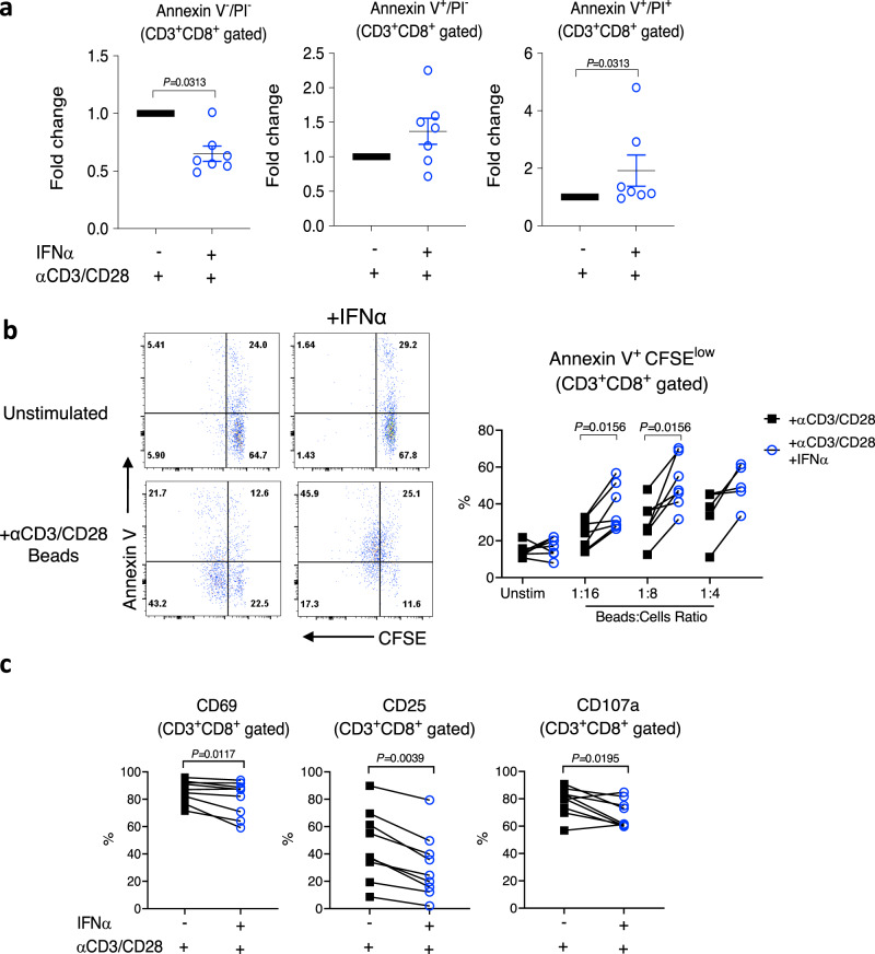
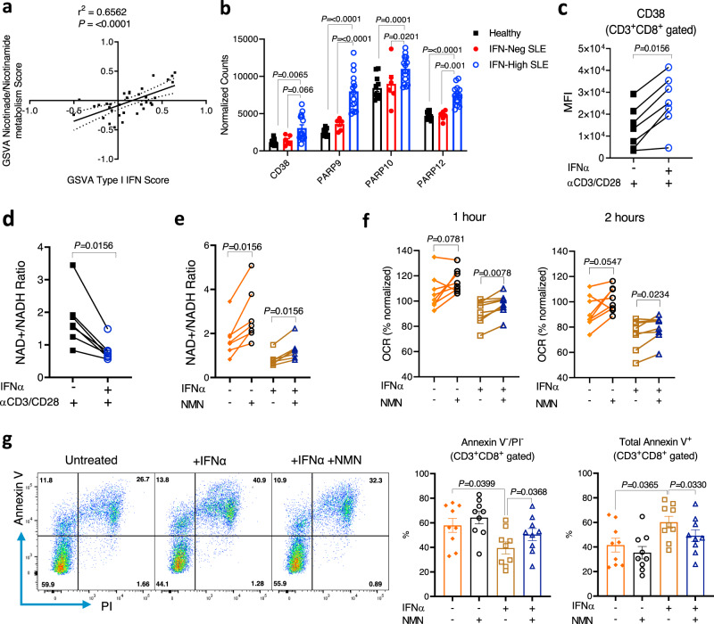
The majority of patients with systemic lupus erythematosus (SLE) have high expression of type I IFN-stimulated genes. Mitochondrial abnormalities have also been reported, but the contribution of type I IFN exposure to these changes is unknown. Here, we show downregulation of mitochondria-derived genes and mitochondria-associated metabolic pathways in IFN-High patients from transcriptomic analysis of CD4 and CD8 T cells. CD8 T cells from these patients have enlarged mitochondria and lower spare respiratory capacity associated with increased cell death upon rechallenge with TCR stimulation. These mitochondrial abnormalities can be phenocopied by exposing CD8 T cells from healthy volunteers to type I IFN and TCR stimulation. Mechanistically these 'SLE-like' conditions increase CD8 T cell NAD+ consumption resulting in impaired mitochondrial respiration and reduced cell viability, both of which can be rectified by NAD+ supplementation. Our data suggest that type I IFN exposure contributes to SLE pathogenesis by promoting CD8 T cell death via metabolic rewiring.
大多数系统性红斑狼疮 (SLE) 患者的 I 型干扰素刺激基因表达水平较高。也有报道称存在线粒体异常,但 I 型干扰素暴露对这些变化的贡献尚不清楚。在这里,我们通过对 CD4 和 CD8 T 细胞的转录组分析显示,IFN-High 患者的线粒体衍生基因和与线粒体相关的代谢途径下调。这些患者的 CD8 T 细胞的线粒体增大,备用呼吸能力降低,与 TCR 刺激后再次受到挑战时细胞死亡增加有关。通过将健康志愿者的 CD8 T 细胞暴露于 I 型干扰素和 TCR 刺激,可以模拟这些“SLE 样”条件下的线粒体异常。从机制上讲,这些“SLE 样”条件通过增加 CD8 T 细胞 NAD+消耗,导致线粒体呼吸受损和细胞活力降低,而 NAD+补充可以纠正这两种情况。我们的数据表明,I 型干扰素暴露通过代谢重编程促进 CD8 T 细胞死亡,从而导致 SLE 的发病机制。