Chen Hui, Li Zhao, Feng Sheng, Wang Anni, Richard-Greenblatt Melissa, Hutson Emily, Andrianus Stefen, Glaser Laurel J, Rodino Kyle G, Qian Jianing, Jayaraman Dinesh, Collman Ronald G, Glascock Abigail, Bushman Frederic D, Lee Jae Seung, Cherry Sara, Fausto Alejandra, Weiss Susan R, Koo Hyun, Corby Patricia M, O'Doherty Una, Garfall Alfred L, Vogl Dan T, Stadtmauer Edward A, Wang Ping
Department of Pathology and Laboratory Medicine, University of Pennsylvania, Philadelphia, PA.
Bioengineering Graduate Program, University of Pennsylvania, Philadelphia, PA.
medRxiv. 2021 Mar 26:2021.03.17.21253847. doi: 10.1101/2021.03.17.21253847.
Little is known about the dynamics of SARS-CoV-2 antigen burden in respiratory samples in different patient populations at different stages of infection. Current rapid antigen tests cannot quantitate and track antigen dynamics with high sensitivity and specificity in respiratory samples.
We developed and validated an ultra-sensitive SARS-CoV-2 antigen assay with smartphone readout using the Microbubbling Digital Assay previously developed by our group, which is a platform that enables highly sensitive detection and quantitation of protein biomarkers. A computer vision-based algorithm was developed for microbubble smartphone image recognition and quantitation. A machine learning-based classifier was developed to classify the smartphone images based on detected microbubbles. Using this assay, we tracked antigen dynamics in serial swab samples from COVID patients hospitalized in ICU and immunocompromised COVID patients.
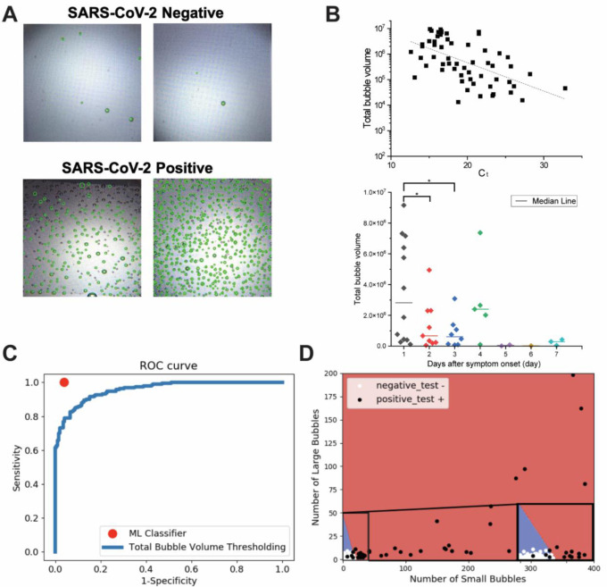
The limit of detection (LOD) of the Microbubbling SARS-CoV-2 Antigen Assay was 0.5 pg/mL (10.6 fM) recombinant nucleocapsid (N) antigen or 4000 copies/mL inactivated SARS-CoV-2 virus in nasopharyngeal (NP) swabs, comparable to many rRT-PCR methods. The assay had high analytical specificity towards SARS-CoV-2. Compared to EUA-approved rRT-PCR methods, the Microbubbling Antigen Assay demonstrated a positive percent agreement (PPA) of 97% (95% confidence interval (CI), 92-99%) in symptomatic individuals within 7 days of symptom onset and positive SARS-CoV-2 nucleic acid results, and a negative percent agreement (NPA) of 97% (95% CI, 94-100%) in symptomatic and asymptomatic individuals with negative nucleic acid results. Antigen positivity rate in NP swabs gradually decreased as days-after-symptom-onset increased, despite persistent nucleic acid positivity of the same samples. The computer vision and machine learning-based automatic microbubble image classifier could accurately identify positives and negatives, based on microbubble counts and sizes. Total microbubble volume, a potential marker of antigen burden, correlated inversely with Ct values and days-after-symptom-onset. Antigen was detected for longer periods of time in immunocompromised patients with hematologic malignancies, compared to immunocompetent individuals. Simultaneous detectable antigens and nucleic acids may indicate the presence of replicating viruses in patients with persistent infections.
The Microbubbling SARS-CoV-2 Antigen Assay enables sensitive and specific detection of acute infections, and quantitation and tracking of antigen dynamics in different patient populations at various stages of infection. With smartphone compatibility and automated image processing, the assay is well-positioned to be adapted for point-of-care diagnosis and to explore the clinical implications of antigen dynamics in future studies.
对于严重急性呼吸综合征冠状病毒2(SARS-CoV-2)抗原负荷在不同感染阶段不同患者群体呼吸道样本中的动态变化,人们了解甚少。目前的快速抗原检测无法在呼吸道样本中以高灵敏度和特异性定量并追踪抗原动态变化。
我们利用本团队之前开发的微泡数字检测法,开发并验证了一种可通过智能手机读取结果的超灵敏SARS-CoV-2抗原检测方法,该平台能够对蛋白质生物标志物进行高灵敏度检测和定量。开发了一种基于计算机视觉的算法用于微泡智能手机图像识别和定量。开发了一种基于机器学习的分类器,根据检测到的微泡对智能手机图像进行分类。使用该检测方法,我们追踪了入住重症监护病房(ICU)的新冠肺炎患者和免疫功能低下的新冠肺炎患者连续拭子样本中的抗原动态变化。
微泡SARS-CoV-2抗原检测法的检测限为0.5 pg/mL(10.6 fM)重组核衣壳(N)抗原,或鼻咽拭子中4000拷贝/mL的灭活SARS-CoV-2病毒,与许多逆转录聚合酶链反应(rRT-PCR)方法相当。该检测方法对SARS-CoV-2具有高分析特异性。与经紧急使用授权(EUA)批准的rRT-PCR方法相比,微泡抗原检测法在症状出现7天内且SARS-CoV-2核酸检测结果为阳性的有症状个体中,阳性百分一致性(PPA)为97%(95%置信区间(CI),92-99%);在核酸检测结果为阴性的有症状和无症状个体中,阴性百分一致性(NPA)为97%(95%CI,94-100%)。尽管相同样本的核酸持续呈阳性,但随着症状出现后天数增加,鼻咽拭子中的抗原阳性率逐渐下降。基于计算机视觉和机器学习的自动微泡图像分类器可根据微泡数量和大小准确识别阳性和阴性。总微泡体积作为抗原负荷的一个潜在标志物,与Ct值和症状出现后天数呈负相关。与免疫功能正常的个体相比,血液系统恶性肿瘤免疫功能低下的患者中抗原检测时间更长。同时检测到抗原和核酸可能表明持续性感染患者中存在正在复制的病毒。
微泡SARS-CoV-2抗原检测法能够灵敏且特异地检测急性感染,并对不同感染阶段不同患者群体的抗原动态变化进行定量和追踪。由于与智能手机兼容且具有自动化图像处理功能,该检测方法很适合用于即时诊断,并在未来研究中探索抗原动态变化的临床意义。