• 文献检索
  • 文档翻译
  • 深度研究
  • 学术资讯
  • Suppr Zotero 插件Zotero 插件
  • 邀请有礼
  • 套餐&价格
  • 历史记录
应用&插件
Suppr Zotero 插件Zotero 插件浏览器插件Mac 客户端Windows 客户端微信小程序
定价
高级版会员购买积分包购买API积分包
服务
文献检索文档翻译深度研究API 文档MCP 服务
关于我们
关于 Suppr公司介绍联系我们用户协议隐私条款
关注我们

Suppr 超能文献

核心技术专利:CN118964589B侵权必究
粤ICP备2023148730 号-1Suppr @ 2026

文献检索

告别复杂PubMed语法,用中文像聊天一样搜索,搜遍4000万医学文献。AI智能推荐,让科研检索更轻松。

立即免费搜索

文件翻译

保留排版,准确专业,支持PDF/Word/PPT等文件格式,支持 12+语言互译。

免费翻译文档

深度研究

AI帮你快速写综述,25分钟生成高质量综述,智能提取关键信息,辅助科研写作。

立即免费体验

在拟行种植体部位,发现牙内吸收比其他阻射性病变更多。

Endodontic remnants are found more than other radiopacities in proposed implant sites.

机构信息

Oral and Maxillofacial Surgery Department, Faculty of Dentistry, Mansoura University, 60 Elgomhoria Street, Mansoura, 35516, Egypt.

Conservative Dentistry Department, Faculty of Dentistry, Zagazig University, Zagazig, Egypt.

出版信息

Int J Implant Dent. 2021 Apr 2;7(1):33. doi: 10.1186/s40729-021-00307-0.

DOI:10.1186/s40729-021-00307-0
PMID:33796914
原文链接:https://pmc.ncbi.nlm.nih.gov/articles/PMC8017025/
Abstract

BACKGROUND

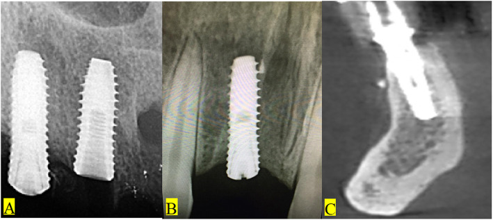
Foreign bodies may be a cause of concern in dental implant failure.

PURPOSE

The aim of the present study was to assess the occurrence and to evaluate the types of radiopacities in dental extraction sites using cone beam computed tomography (CBCT).

MATERIALS AND METHODS

The incidence, location, and types of radiopacities were evaluated in 180 CBCT scans.

RESULTS

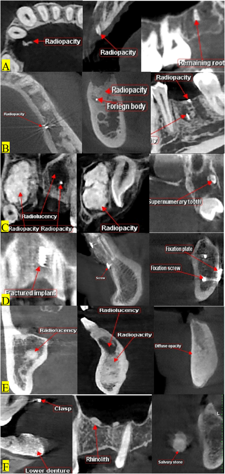
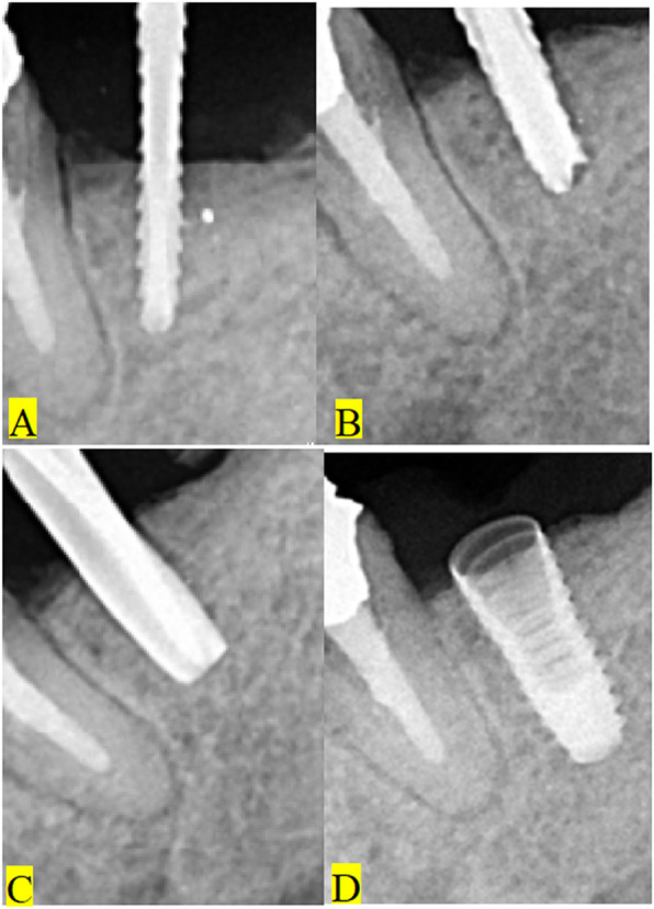
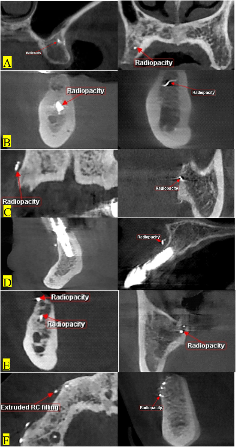
Different radiopaque structures could be noted in 84 scans. Foreign bodies and remaining roots were frequently seen. Most of the radiopacities were attributed to remaining endodontic filling in upper and lower jaws in 25 scans in different locations. Remaining roots could be detected in 20 scans. Focal and diffuse radiopaque bony lesions were observed in 16 scans. Tissue response in the form of radiolucency could be seen more with endodontic foreign bodies. Tissue reactions to radiopaque filling remnants were seen in 6.11% of cases.

CONCLUSIONS

Foreign body remnants, mostly of endodontic fillings, were frequently seen in CBCT in upper and lower jaws. Evidence of tissue reactions to extraction remnants could be found. Endodontic filling remnants could be seen more in the upper jaw. Thorough examination of implant site for the presence of endodontic foreign body remnants should be stressed. Debridement of the extraction socket should be done carefully in endodontically treated teeth.

摘要

背景

异物可能是导致牙种植体失败的一个原因。

目的

本研究旨在通过锥形束 CT(CBCT)评估拔牙部位的放射性不透射线体的发生和类型。

材料和方法

对 180 例 CBCT 扫描的发生率、位置和类型进行了评估。

结果

在 84 个扫描中可以观察到不同的不透射线结构。经常可见异物和残根。在不同部位的 25 个扫描中,大多数放射性不透射线体归因于上颌和下颌的残留牙髓填充物。在 20 个扫描中可以检测到残留的牙根。在 16 个扫描中观察到局灶性和弥漫性放射性不透射线骨病变。在有牙髓异物的情况下,更常见到以射线透明为特征的组织反应。在 6.11%的病例中观察到对放射性不透射线填充物残余的组织反应。

结论

在上颌和下颌的 CBCT 中经常可以看到异物残体,主要是牙髓填充物。可以发现对拔牙残体的组织反应的证据。在上颌中可以看到更多的牙髓填充物残余。应该强调在种植体部位彻底检查是否存在牙髓异物残体。对于接受过牙髓治疗的牙齿,应仔细清创拔牙窝。

https://cdn.ncbi.nlm.nih.gov/pmc/blobs/0215/8017025/4e3e06296996/40729_2021_307_Fig5_HTML.jpg
https://cdn.ncbi.nlm.nih.gov/pmc/blobs/0215/8017025/331ec6b4cd47/40729_2021_307_Fig1_HTML.jpg
https://cdn.ncbi.nlm.nih.gov/pmc/blobs/0215/8017025/50436a9138eb/40729_2021_307_Fig2_HTML.jpg
https://cdn.ncbi.nlm.nih.gov/pmc/blobs/0215/8017025/294c6d82dd9e/40729_2021_307_Fig3_HTML.jpg
https://cdn.ncbi.nlm.nih.gov/pmc/blobs/0215/8017025/5ee1ee9516a6/40729_2021_307_Fig4_HTML.jpg
https://cdn.ncbi.nlm.nih.gov/pmc/blobs/0215/8017025/4e3e06296996/40729_2021_307_Fig5_HTML.jpg

相似文献

1
Endodontic remnants are found more than other radiopacities in proposed implant sites.在拟行种植体部位,发现牙内吸收比其他阻射性病变更多。
Int J Implant Dent. 2021 Apr 2;7(1):33. doi: 10.1186/s40729-021-00307-0.
2
Endodontic outcome predictors identified with periapical radiographs and cone-beam computed tomography scans.利用根尖放射线片和锥形束 CT 扫描识别牙髓病治疗结果的预测因素。
J Endod. 2011 Mar;37(3):326-31. doi: 10.1016/j.joen.2010.11.032.
3
Detection of Fractured Endodontic Instruments in Root Canals: Comparison between Different Digital Radiography Systems and Cone-beam Computed Tomography.根管内折断牙髓治疗器械的检测:不同数字放射成像系统与锥形束计算机断层扫描的比较
J Endod. 2017 Apr;43(4):544-549. doi: 10.1016/j.joen.2016.11.017. Epub 2017 Feb 16.
4
Assessment of periapical health, quality of root canal filling, and coronal restoration by using cone-beam computed tomography.使用锥形束计算机断层扫描评估根尖周健康状况、根管充填质量和冠部修复情况。
Niger J Clin Pract. 2016 Sep-Oct;19(5):673-7. doi: 10.4103/1119-3077.188697.
5
Influence of Endodontic Treatment and Coronal Restoration on Status of Periapical Tissues: A Cone-beam Computed Tomographic Study.根管治疗和冠部修复对根尖周组织状况的影响:一项锥形束计算机断层扫描研究。
J Endod. 2015 Oct;41(10):1614-8. doi: 10.1016/j.joen.2015.07.008. Epub 2015 Aug 22.
6
Quality of Endodontic Treatment and Prevalence of Apical Radiolucencies in a Bulgarian Subpopulation: a CBCT Analysis.保加利亚亚人群的根管治疗质量和根尖透射线的流行率:CBCT 分析。
Folia Med (Plovdiv). 2021 Feb 28;63(1):81-87. doi: 10.3897/folmed.63.e52204.
7
Volumetric changes in apical radiolucencies of endodontically treated teeth assessed by cone-beam computed tomography 1 year after orthograde retreatment.通过锥形束计算机断层扫描评估根管再治疗1年后根管治疗牙齿根尖部透射区的体积变化。
J Endod. 2013 Dec;39(12):1504-9. doi: 10.1016/j.joen.2013.08.034. Epub 2013 Oct 15.
8
Prevalence of Apical Bone Defects and Evaluation of Associated Factors Detected with Cone-beam Computed Tomographic Images.根尖骨缺损的患病率及锥束计算机断层扫描图像检测相关因素的评估
J Endod. 2015 Jul;41(7):1043-7. doi: 10.1016/j.joen.2015.03.011. Epub 2015 Apr 25.
9
Unexpected radiopaque foreign bodies encountered in dental practice.牙科诊疗中意外发现的不透射线异物。
Quintessence Int. 2019;50(2):146-155. doi: 10.3290/j.qi.a41707.
10
Detection of vertical root fractures by conventional radiographic examination and cone beam computed tomography - an in vitro analysis.常规放射学检查和锥形束计算机断层扫描检测垂直根折:一项体外分析。
Dent Traumatol. 2013 Feb;29(1):41-6. doi: 10.1111/j.1600-9657.2012.01126.x. Epub 2012 Mar 13.

引用本文的文献

1
Physicochemical properties and biological interaction of calcium silicate-based sealers - in vivo model.硅酸钙基封闭剂的物理化学性质及生物相互作用——体内模型
Clin Oral Investig. 2025 Jan 25;29(1):86. doi: 10.1007/s00784-025-06150-6.

本文引用的文献

1
Prevalence and treatment of retrograde peri-implantitis: a retrospective cohort study covering a 20-year period.逆行性种植体周围炎的流行情况和治疗:一项涵盖 20 年的回顾性队列研究。
Clin Oral Investig. 2021 Jul;25(7):4553-4561. doi: 10.1007/s00784-020-03769-5. Epub 2021 Jan 14.
2
Knowledge and Attitude towards Retrograde Peri-Implantitis among Italian Implantologists: A Cross-Sectional Survey.意大利种植学家对逆行性种植体周围炎的认知和态度:一项横断面调查。
Int J Environ Res Public Health. 2020 Nov 12;17(22):8356. doi: 10.3390/ijerph17228356.
3
Correlation between endodontic pulpal/periapical disease and retrograde peri-implantitis: A case series.
牙髓/根尖周病与逆行性种植体周围炎的相关性:病例系列研究。
Aust Endod J. 2021 Aug;47(2):358-364. doi: 10.1111/aej.12458. Epub 2020 Nov 7.
4
Retrospective cohort study of a tapered implant with high primary stability in patients with local and systemic risk factors-7-year data.对具有局部和全身风险因素的患者使用具有高初始稳定性的锥形种植体的回顾性队列研究——7年数据
Int J Implant Dent. 2018 Dec 17;4(1):41. doi: 10.1186/s40729-018-0151-5.
5
Multiple calcifying hyperplastic dental follicles.多个钙化增生性牙滤泡
J Stomatol Oral Maxillofac Surg. 2019 Feb;120(1):77-79. doi: 10.1016/j.jormas.2018.08.007. Epub 2018 Sep 8.
6
Endodontic-related inferior alveolar nerve injuries: A review and a therapeutic flow chart.牙髓相关的下牙槽神经损伤:综述及治疗流程图。
J Stomatol Oral Maxillofac Surg. 2018 Nov;119(5):412-418. doi: 10.1016/j.jormas.2018.04.012. Epub 2018 May 3.
7
Implant failure and history of failed endodontic treatment: A retrospective case-control study.种植体失败与根管治疗失败史:一项回顾性病例对照研究。
J Clin Exp Dent. 2017 Nov 1;9(11):e1322-e1328. doi: 10.4317/jced.54277. eCollection 2017 Nov.
8
Factors affecting the periapical healing process of endodontically treated teeth.影响根管治疗后牙齿根尖周愈合过程的因素。
J Appl Oral Sci. 2017 Sep-Oct;25(5):465-476. doi: 10.1590/1678-7757-2016-0464.
9
Periodontal and endodontic pathology delays extraction socket healing in a canine model.在犬类模型中,牙周和牙髓病理学延迟拔牙创口愈合。
J Periodontal Implant Sci. 2017 Jun;47(3):143-153. doi: 10.5051/jpis.2017.47.3.143. Epub 2017 Jun 28.
10
Apical/Retrograde Periimplantitis/Implant Periapical Lesion: Etiology, Risk Factors, and Treatment Options: A Systematic Review.根尖/逆行性种植体周围炎/种植体根尖周病变:病因、危险因素及治疗选择:一项系统评价
Implant Dent. 2016 Oct;25(5):684-97. doi: 10.1097/ID.0000000000000424.