Department of Pediatric Surgery, Second Affiliated Hospital of Xi'an Jiaotong University, Xi'an, 710004 Shaanxi, China; Department of Biomedical Engineering, Pratt School of Engineering, Duke University, Durham, NC 27708, USA.
Department of Biomedical Engineering, Pratt School of Engineering, Duke University, Durham, NC 27708, USA.
Cell Stem Cell. 2021 Apr 1;28(4):603-622. doi: 10.1016/j.stem.2021.02.010.
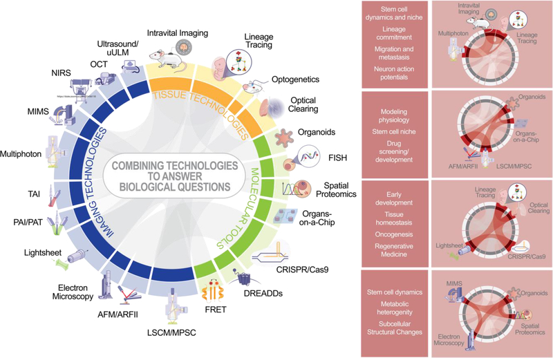
What you see is what you get-imaging techniques have long been essential for visualization and understanding of tissue development, homeostasis, and regeneration, which are driven by stem cell self-renewal and differentiation. Advances in molecular and tissue modeling techniques in the last decade are providing new imaging modalities to explore tissue heterogeneity and plasticity. Here we describe current state-of-the-art imaging modalities for tissue research at multiple scales, with a focus on explaining key tradeoffs such as spatial resolution, penetration depth, capture time/frequency, and moieties. We explore emerging tissue modeling and molecular tools that improve resolution, specificity, and throughput.
所见即所得——成像技术长期以来一直是组织发育、稳态和再生的可视化和理解的关键,而这些过程是由干细胞自我更新和分化所驱动的。在过去十年中,分子和组织建模技术的进步为探索组织异质性和可塑性提供了新的成像方式。在这里,我们描述了用于多尺度组织研究的当前最先进的成像方式,重点解释了空间分辨率、穿透深度、捕获时间/频率和部分等关键权衡。我们还探讨了提高分辨率、特异性和通量的新兴组织建模和分子工具。