Simeoni Francesco, Del Signore Francesca, Aste Giovanni, Bargellini Paolo, Rubini Giuseppe, Terragni Rossella, Tamburro Roberto, Falerno Ilaria, de Pasquale Francesco, Russo Marco, Vignoli Massimo
Faculty of Veterinary Medicine, University of Teramo, SP 18, 64100 Teramo, Italy.
Tyrus Veterinary Clinic, via Aldo Bartocci 1G, 05100 Terni, Italy.
Animals (Basel). 2021 Mar 3;11(3):670. doi: 10.3390/ani11030670.
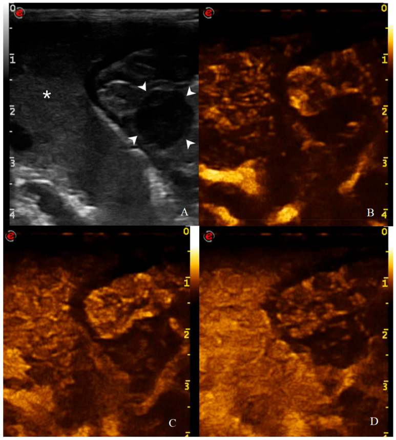
Canine gastric disorders are common in veterinary clinical practice and among these neoplasms require rapid identification and characterization. Standard ultrasound (US) is the imaging modality of choice for gastric wall assessment. The aim of this prospective study is to describe the specific B-mode and contrast enhanced US (CEUS) features of normal, inflammatory, and neoplastic gastric wall in dogs. B-mode US and CEUS of the stomach were performed in anesthetized dogs with or without gastric disorders. Gastric wall qualitative and quantitative parameters were evaluated on B-mode US and CEUS examination. A total of 41 dogs were included: 6 healthy (HEA) as the control group; 9 gastritis (INF); 8 adenocarcinoma (AC); 8 alimentary lymphoma (AL); 4 leiomyosarcoma (LEIS); 2 gastrointestinal stromal tumor (GIST); 2 leiomyoma; 1 undifferentiated sarcoma; 1 metastatic gastric hemangiosarcoma. Gastric tumors appear as a marked wall thickness with absent layers definition and possible regional lymphadenopathy (AC and AL) and steatitis (AC) while gastritis generally shows no/mild thickening and no other alterations on B-mode US. On CEUS, neoplasm shows a higher and faster wash in if compared to that of gastritis. B-mode and CEUS assessment may be useful in the evaluation of canine gastric disorders in the distinction between gastritis and gastric neoplasms, even if there are no specific features able to discriminate between the different tumor histotypes.
犬胃部疾病在兽医临床实践中很常见,其中肿瘤需要快速识别和特征描述。标准超声(US)是评估胃壁的首选成像方式。这项前瞻性研究的目的是描述犬正常、炎症性和肿瘤性胃壁的特定B模式和对比增强超声(CEUS)特征。对有或无胃部疾病的麻醉犬进行胃的B模式超声和CEUS检查。在B模式超声和CEUS检查中评估胃壁的定性和定量参数。共纳入41只犬:6只健康犬(HEA)作为对照组;9只患胃炎(INF);8只患腺癌(AC);8只患消化道淋巴瘤(AL);4只患平滑肌肉瘤(LEIS);2只患胃肠道间质瘤(GIST);2只患平滑肌瘤;1只患未分化肉瘤;1只患转移性胃血管肉瘤。胃肿瘤表现为胃壁明显增厚,层次不清,可能伴有区域淋巴结病(AC和AL)和脂肪炎(AC),而胃炎在B模式超声上通常显示无增厚/轻度增厚且无其他改变。在CEUS上,与胃炎相比,肿瘤显示出更高、更快的早期增强。B模式和CEUS评估在区分胃炎和胃肿瘤的犬胃部疾病评估中可能有用,即使没有能够区分不同肿瘤组织学类型的特异性特征。