Institute of Anatomy and Cell Biology, Julius-Maximilians-University Würzburg, 97070 Würzburg, Germany.
Institute of Experimental Biomedicine, Chair I, University Hospital Würzburg, 97080 Würzburg, Germany.
Cells. 2021 Mar 24;10(4):722. doi: 10.3390/cells10040722.
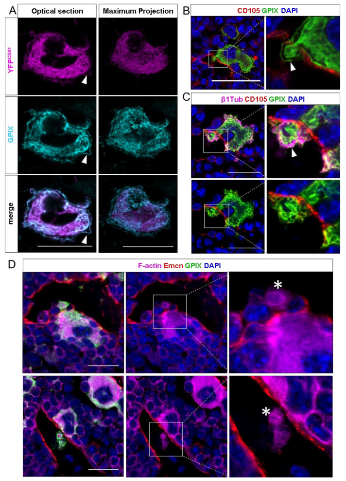
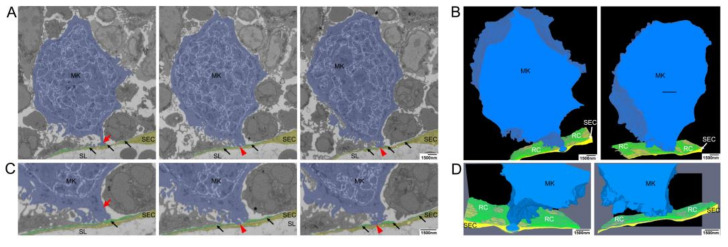
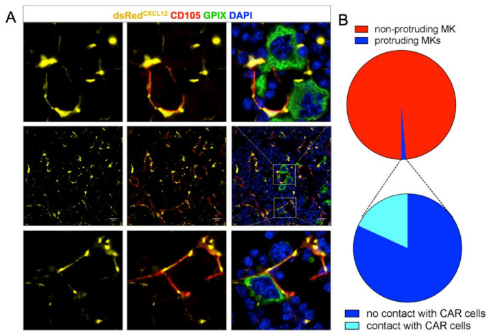
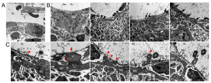
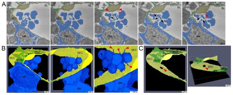
Megakaryocytes (MKs) release platelets into the lumen of bone marrow (BM) sinusoids while remaining to reside within the BM. The morphogenetic events of this complex process are still not fully understood. We combined confocal laser scanning microscopy with transmission and serial block-face scanning electron microscopy followed by 3D-reconstruction on mouse BM tissue sections. These analyses revealed that MKs in close vicinity to BM sinusoid (BMS) wall first induce the lateral retraction of CXCL12-abundant reticular (CAR) cells (CAR), followed by basal lamina (BL) degradation enabling direct MK-sinusoidal endothelial cells (SECs) interaction. Subsequently, an endothelial engulfment starts that contains a large MK protrusion. Then, MK protrusions penetrate the SEC, transmigrate into the BMS lumen and form proplatelets that are in direct contact to the SEC surface. Furthermore, such processes are induced on several sites, as observed by 3D reconstructions. Our data demonstrate that MKs in interaction with CAR-cells actively induce BMS wall alterations, including CAR-cell retraction, BL degradation, and SEC engulfment containing a large MK protrusion. This results in SEC penetration enabling the migration of MK protrusion into the BMS lumen where proplatelets that are adherent to the luminal SEC surface are formed and contribute to platelet release into the blood circulation.
巨核细胞 (MKs) 在骨髓 (BM) 窦状隙的管腔中释放血小板,同时仍驻留在 BM 中。这个复杂过程的形态发生事件仍未完全了解。我们将共聚焦激光扫描显微镜与透射和连续块面扫描电子显微镜相结合,对小鼠 BM 组织切片进行 3D 重建。这些分析表明,靠近 BM 窦壁的 MKs 首先诱导 CXCL12 丰富的网状 (CAR) 细胞 (CAR) 的侧向回缩,随后基底膜 (BL) 降解,使 MK 与窦内皮细胞 (SEC) 直接相互作用。随后,开始发生内皮细胞吞噬作用,其中包含一个大的 MK 突起。然后,MK 突起穿透 SEC,迁移到 BMS 管腔中,并形成与 SEC 表面直接接触的原血小板。此外,通过 3D 重建观察到,这种过程发生在几个部位。我们的数据表明,与 CAR 细胞相互作用的 MKs 主动诱导 BMS 壁的改变,包括 CAR 细胞回缩、BL 降解和包含大 MK 突起的 SEC 吞噬作用。这导致 SEC 穿透,允许 MK 突起迁移到 BMS 管腔中,在那里形成与管腔 SEC 表面黏附的原血小板,并有助于血小板释放到血液循环中。