Cellular Biology Laboratory, Department of Research in Pulmonary Fibrosis, Instituto Nacional de Enfermedades Respiratorias Ismael Cosío Villegas (INER), Mexico City 14080, Mexico.
Laboratory of Pulmonary Biopathology INER-Ciencias UNAM, Department of Research in Pulmonary Fibrosis, Instituto Nacional de Enfermedades Respiratorias Ismael Cosío Villegas (INER), Mexico City 14080, Mexico.
Biomolecules. 2021 Mar 4;11(3):378. doi: 10.3390/biom11030378.
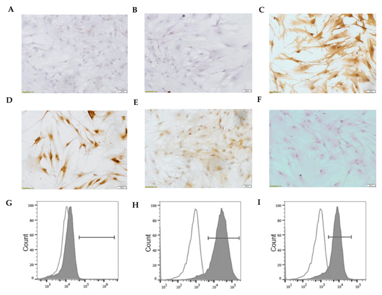
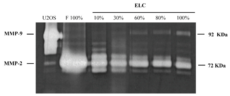
. In passages above ten and growing very actively, we observed that some human lung fibroblasts cultured under standard conditions were transformed into a lineage of epithelial-like cells (ELC). To systematically evaluate the possible mesenchymal-epithelial transition (MET) occurrence, fibroblasts were obtained from normal lungs and also from lungs affected by idiopathic interstitial diseases. When an unusual epithelial-like phenotypic change was observed, cultured cells were characterized by confocal immunofluorescence microscopy, immunoblotting, immunocytochemistry, cytofluorometry, gelatin zymography, RT-qPCR, and hybridization in a whole-transcript human microarray. Additionally, microvesicles fraction (MVs) from ELC and fibroblasts were used to induce MET, while the microRNAs (miRNAs) contained in the MVs were identified. Pattern-gene expression of the original fibroblasts and the derived ELC revealed profound changes, upregulating characteristic epithelial-cell genes and downregulating mesenchymal genes, with a marked increase of E-cadherin, cytokeratin, and ZO-1, and the loss of expression of α-SMA, collagen type I, and Thy-1 cell surface antigen (CD90). Fibroblasts, exposed to culture media or MVs from the ELC, acquired ELC phenotype. The miRNAs in MVs shown six expressed exclusively in fibroblasts, and three only in ELC; moreover, twelve miRNAs were differentially expressed between fibroblasts and ELC, all of them but one was overexpressed in fibroblasts. These findings suggest that the MET-like process can occur in human lung fibroblasts, either from normal or diseased lungs. However, the biological implication is unclear.
. 在长度超过十且生长非常活跃的段落中,我们观察到,在标准条件下培养的一些人肺成纤维细胞被转化为上皮样细胞系(ELC)。为了系统地评估可能发生的间充质上皮转化(MET),我们从正常肺和特发性间质性疾病肺中获得成纤维细胞。当观察到不寻常的上皮样表型变化时,通过共聚焦免疫荧光显微镜、免疫印迹、免疫细胞化学、细胞荧光计、明胶酶谱、RT-qPCR 和全转录人微阵列杂交对培养细胞进行了特征描述。此外,使用 ELC 和成纤维细胞的微泡(MVs)来诱导 MET,同时鉴定 MVs 中包含的 microRNAs(miRNAs)。原始成纤维细胞和衍生的 ELC 的模式基因表达显示出深刻的变化,上调特征上皮细胞基因并下调间充质基因,E-钙粘蛋白、细胞角蛋白和 ZO-1 的表达明显增加,而α-SMA、I 型胶原和 Thy-1 细胞表面抗原(CD90)的表达减少。暴露于培养基或 ELC 的 MVs 的成纤维细胞获得 ELC 表型。MVs 中的 miRNAs 显示有六个仅在成纤维细胞中表达,三个仅在 ELC 中表达;此外,在成纤维细胞和 ELC 之间有 12 个 miRNAs 差异表达,其中除一个外均在成纤维细胞中过表达。这些发现表明,MET 样过程可以在人肺成纤维细胞中发生,无论是来自正常肺还是疾病肺。然而,其生物学意义尚不清楚。