Kedra Alice, Dohan Anthony, Gaujoux Sébastien, Sibony Mathilde, Jouinot Anne, Assié Guillaume, Groussin Rouiller Lionel, Libé Rossella, Bertherat Jérôme, Soyer Philippe, Barat Maxime
Department of Diagnostic and Interventional Imaging, Hôpital Cochin, Assistance Publique-Hôpitaux de Paris, 75014 Paris, France.
Faculté de Médecine, Université de Paris, 75006 Paris, France.
Cancers (Basel). 2021 Mar 31;13(7):1603. doi: 10.3390/cancers13071603.
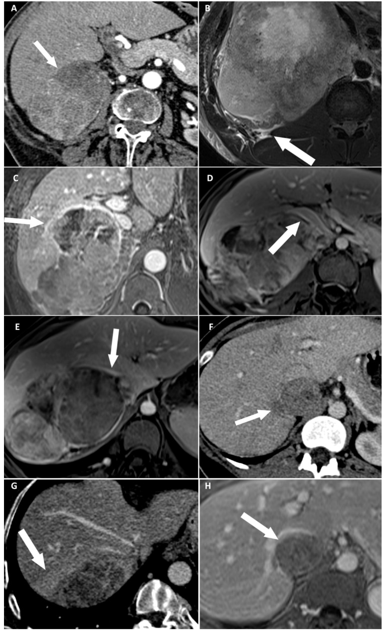
The major prognosis factor of adrenocortical carcinoma (ACC) is the completeness of surgery. The aim of our study was to identify preoperative imaging features associated with direct liver involvement (DLI) by right-sided ACC. Two radiologists, blinded to the outcome, independently reviewed preoperative CT and MRI examinations for eight signs of DLI, in patients operated for right-sided ACC and retrospectively included from November 2007 to January 2020. DLI was confirmed using surgical and histopathological findings. Kappa values were calculated. Univariable and multivariable analyses were performed by using a logistic regression model. Receiver operating characteristic (ROC) curves were built for CT and MRI. Twenty-nine patients were included. Seven patients had DLI requiring resection. At multivariable analysis, focal ACC bulge was the single independent sign associated with DLI on CT (OR: 60.00; 95% CI: 4.60-782.40; < 0.001), and ACC contour disruption was the single independent sign associated with DLI on MRI (OR: 126.00; 95% CI: 6.82-2328.21; < 0.001). Both signs were highly reproducible, with respective kappa values of 0.85 and 0.91. The areas under ROC curves of MRI and CT models were not different ( = 0.838). Focal ACC bulge on CT and ACC contour disruption on MRI are independent and highly reproducible signs, strongly associated with DLI by right-sided ACC on preoperative imaging. MRI does not improve the preoperative assessment of DLI by comparison with CT.
肾上腺皮质癌(ACC)的主要预后因素是手术的完整性。我们研究的目的是确定与右侧ACC直接肝侵犯(DLI)相关的术前影像学特征。两名对结果不知情的放射科医生,独立回顾了2007年11月至2020年1月期间接受右侧ACC手术且回顾性纳入研究的患者的术前CT和MRI检查,以寻找DLI的八个征象。通过手术和组织病理学结果确认DLI。计算Kappa值。使用逻辑回归模型进行单变量和多变量分析。绘制CT和MRI的受试者操作特征(ROC)曲线。共纳入29例患者。7例患者有DLI需要切除。多变量分析显示,局灶性ACC隆起是CT上与DLI相关的唯一独立征象(OR:60.00;95%CI:4.60 - 782.40;P < 0.001),而ACC轮廓中断是MRI上与DLI相关的唯一独立征象(OR:126.00;95%CI:6.82 - 2328.21;P < 0.001)。这两个征象的重复性都很高,Kappa值分别为0.85和0.91。MRI和CT模型的ROC曲线下面积无差异(P = 0.838)。CT上的局灶性ACC隆起和MRI上的ACC轮廓中断是独立且重复性高的征象,在术前影像学上与右侧ACC的DLI密切相关。与CT相比,MRI并未改善DLI的术前评估。