Division of Neonatology, Department of Pediatrics, Children's Mercy Hospital, Kansas City, Missouri, USA.
Genomic Medicine Center, Children's Mercy Hospital, Kansas City, Missouri, USA.
JCI Insight. 2021 Apr 8;6(7):134170. doi: 10.1172/jci.insight.134170.
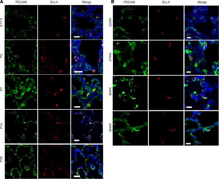
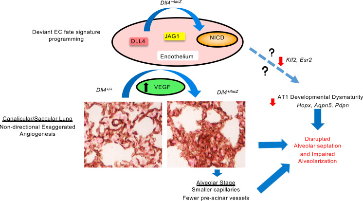
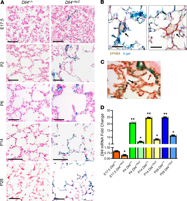
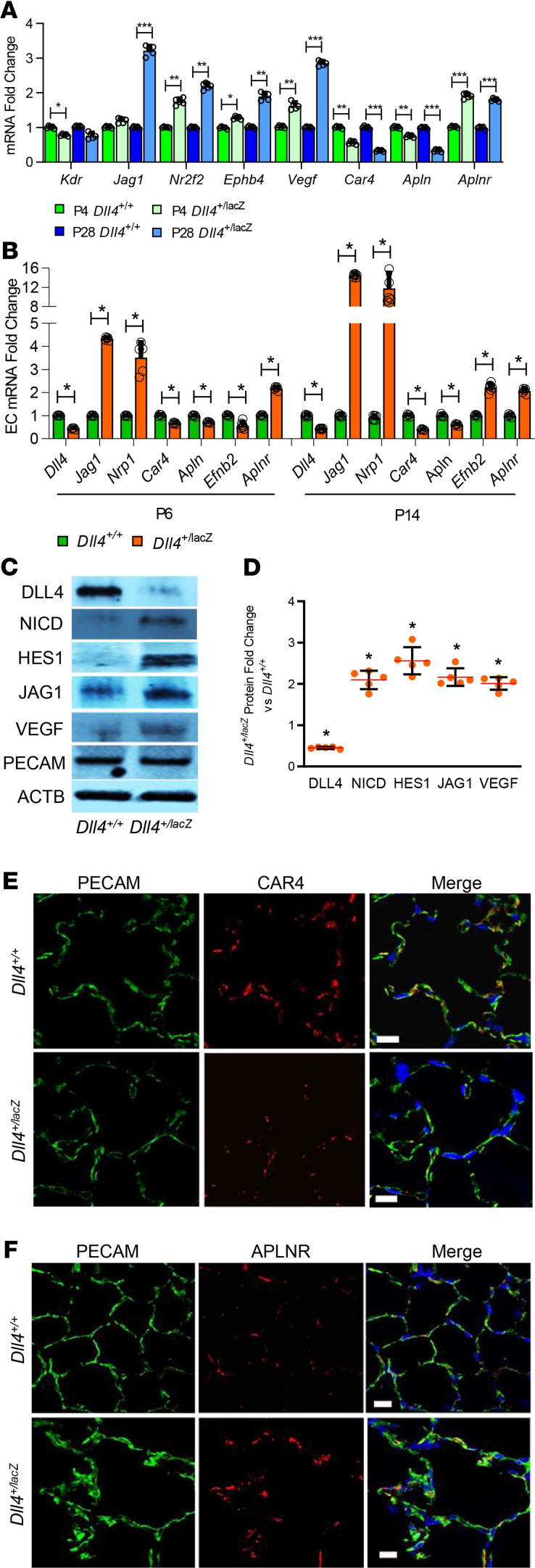
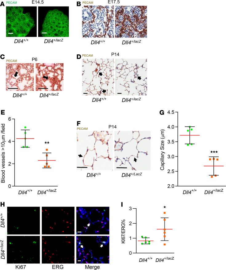
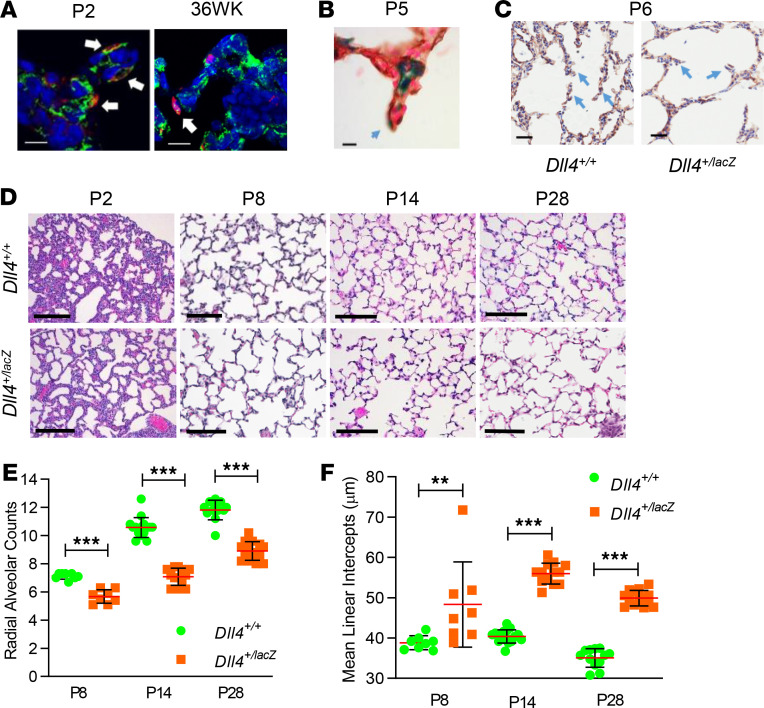
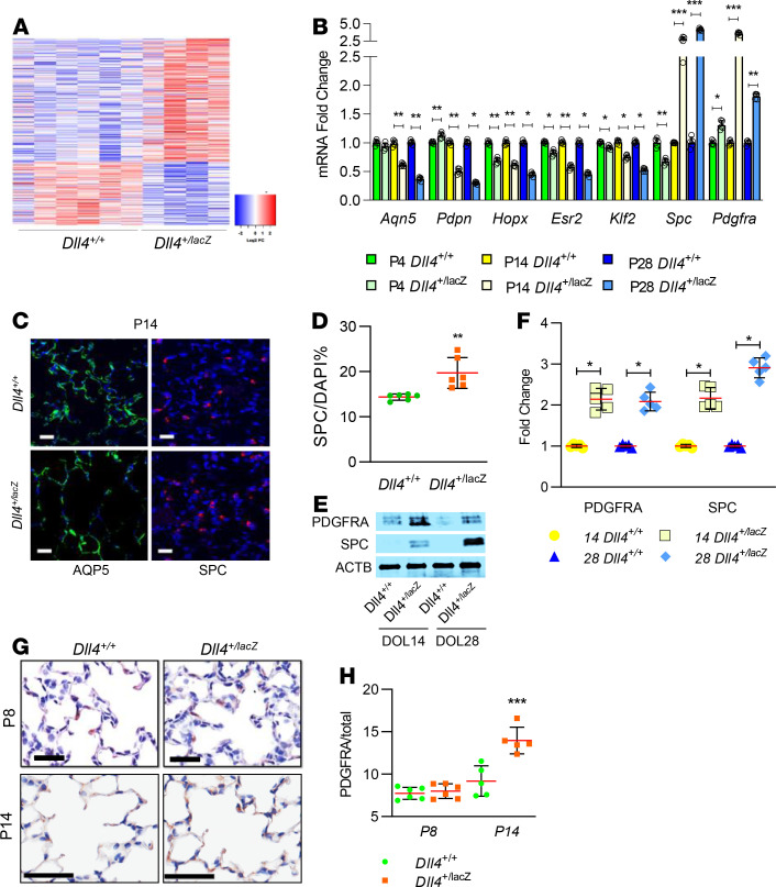
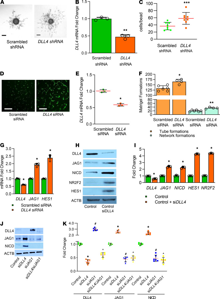
The molecular mechanisms by which endothelial cells (ECs) regulate pulmonary vascularization and contribute to alveolar epithelial cell development during lung morphogenesis remain unknown. We tested the hypothesis that delta-like 4 (DLL4), an EC Notch ligand, is critical for alveolarization by combining lung mapping and functional studies in human tissue and DLL4-haploinsufficient mice (Dll4+/lacz). DLL4 expressed in a PECAM-restricted manner in capillaries, arteries, and the alveolar septum from the canalicular to alveolar stage in mice and humans. Dll4 haploinsufficiency resulted in exuberant, nondirectional vascular patterning at E17.5 and P6, followed by smaller capillaries and fewer intermediate blood vessels at P14. Vascular defects coincided with polarization of lung EC expression toward JAG1-NICD-HES1 signature and decreased tip cell-like (Car4) markers. Dll4+/lacZ mice had impaired terminal bronchiole development at the canalicular stage and impaired alveolarization upon lung maturity. We discovered that alveolar type I cell (Aqp5) markers progressively decreased in Dll4+/lacZ mice after birth. Moreover, in human lung EC, DLL4 deficiency programmed a hypersprouting angiogenic phenotype cell autonomously. In conclusion, DLL4 is expressed from the canalicular to alveolar stage in mice and humans, and Dll4 haploinsufficiency programs dysmorphic microvascularization, impairing alveolarization. Our study reveals an obligate role for DLL4-regulated angiogenesis in distal lung morphogenesis.
内皮细胞 (ECs) 通过何种分子机制调节肺血管生成并促进肺形态发生过程中肺泡上皮细胞的发育尚不清楚。我们通过对人类组织的肺图谱绘制和功能研究,以及 DLL4 部分缺失(Dll4+/lacz)小鼠的实验,检验了内皮 Notch 配体 delta-like 4(DLL4)对于肺泡化至关重要的假说。在从小管期到肺泡期的小鼠和人类肺中,DLL4 以 PECAM 限制的方式在毛细血管、动脉和肺泡隔中表达。Dll4 部分缺失导致 E17.5 和 P6 时出现丰富的、无方向的血管形态发生,随后 P14 时毛细血管较小且中间血管较少。血管缺陷与肺 EC 表达向 JAG1-NICD-HES1 特征的极化以及尖端细胞样(Car4)标志物的减少同时发生。Dll4+/lacZ 小鼠在小管期时出现终末细支气管发育不良,在肺成熟时出现肺泡化受损。我们发现,出生后 Dll4+/lacZ 小鼠的肺泡 I 型细胞(Aqp5)标志物逐渐减少。此外,在人类肺 EC 中,DLL4 缺失自主编程了过度发芽的血管生成表型。总之,DLL4 在小鼠和人类中从小管期到肺泡期表达,Dll4 部分缺失导致畸形的微血管化,从而损害肺泡化。我们的研究揭示了 DLL4 调控的血管生成在远端肺形态发生中的必需作用。