• 文献检索
  • 文档翻译
  • 深度研究
  • 学术资讯
  • Suppr Zotero 插件Zotero 插件
  • 邀请有礼
  • 套餐&价格
  • 历史记录
应用&插件
Suppr Zotero 插件Zotero 插件浏览器插件Mac 客户端Windows 客户端微信小程序
定价
高级版会员购买积分包购买API积分包
服务
文献检索文档翻译深度研究API 文档MCP 服务
关于我们
关于 Suppr公司介绍联系我们用户协议隐私条款
关注我们

Suppr 超能文献

核心技术专利:CN118964589B侵权必究
粤ICP备2023148730 号-1Suppr @ 2026

文献检索

告别复杂PubMed语法,用中文像聊天一样搜索,搜遍4000万医学文献。AI智能推荐,让科研检索更轻松。

立即免费搜索

文件翻译

保留排版,准确专业,支持PDF/Word/PPT等文件格式,支持 12+语言互译。

免费翻译文档

深度研究

AI帮你快速写综述,25分钟生成高质量综述,智能提取关键信息,辅助科研写作。

立即免费体验

脂肪间充质干细胞诱导的 PAX8 通过稳定 TAZ 蛋白促进卵巢癌细胞生长。

Adipose-derived mesenchymal stem cells induced PAX8 promotes ovarian cancer cell growth by stabilizing TAZ protein.

机构信息

Department of Obstetrics, The Affiliated Hospital of Qingdao University, Qingdao, China.

Department of Hepatobiliary and Pancreatic Surgery, The Affiliated Hospital of Qingdao University, Qingdao, China.

出版信息

J Cell Mol Med. 2021 May;25(9):4434-4443. doi: 10.1111/jcmm.16511. Epub 2021 Apr 8.

DOI:10.1111/jcmm.16511
PMID:33830648
原文链接:https://pmc.ncbi.nlm.nih.gov/articles/PMC8093979/
Abstract

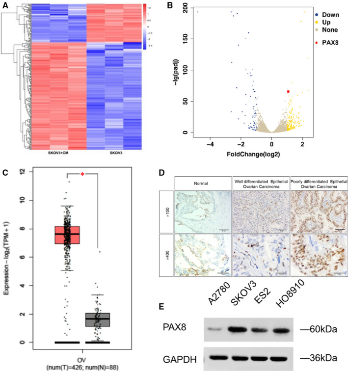
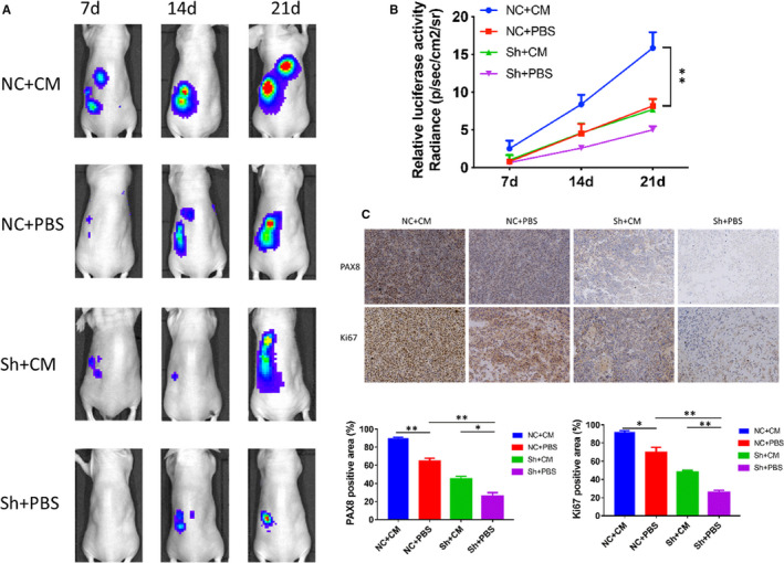
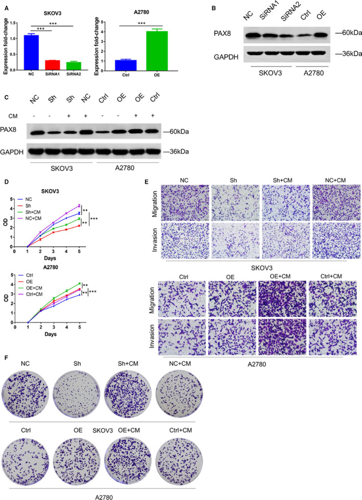
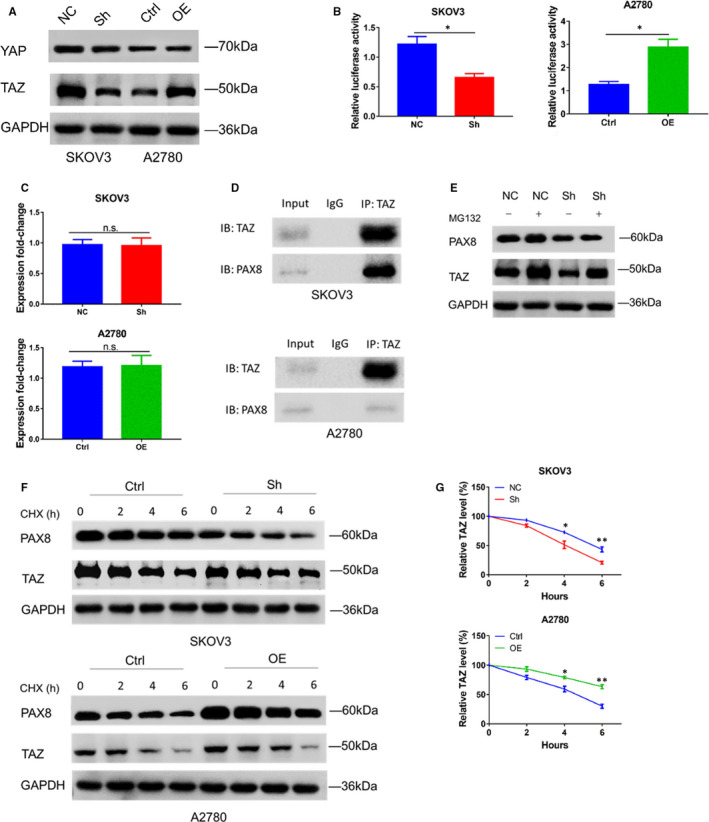
Our previous studies have shown that the Adipose-derived mesenchymal stem cells (ADSCs) can regulate metastasis and development of ovarian cancer. However, its specific mechanism has yet to be fully revealed. In this study, an RNA-seq approach was adopted to compare the differences in mRNA levels in ovarian cancer cells being given or not given ADSCs. The mRNA level of paired box 8 (PAX8) changed significantly and was confirmed as an important factor in tumour-inducing effect of ADSCs. In comparison with the ovarian cancer cells cultured in the common growth medium, those cultured in the medium supplemented with ADSCs showed a significant increase of the PAX8 level. Moreover, the cancer cell growth could be restricted, even in the ADSC-treated group (P < .05), by inhibiting PAX8. In addition, an overexpression of PAX8 could elevate the proliferation of ovarian cancer cells. Moreover, Co-IP assays in ovarian cancer cells revealed that an interaction existed between endogenous PAX8 and TAZ. And the PAX8 levels regulated the degradation of TAZ. The bioluminescence images captured in vivo manifested that the proliferation and the PAX8 expression level in ovarian cancers increased in the ADMSC-treated group, and the effect of ADSCs in promoting tumours was weakened through inhibiting PAX8. Our findings indicate that the PAX8 expression increment could contribute a role in promoting the ADSC-induced ovarian cancer cell proliferation through TAZ stability regulation.

摘要

我们之前的研究表明脂肪间充质干细胞(ADSCs)可以调节卵巢癌细胞的转移和发展。然而,其具体机制尚未完全揭示。在这项研究中,我们采用 RNA-seq 方法比较了给予或未给予 ADSCs 的卵巢癌细胞之间 mRNA 水平的差异。配对盒 8(PAX8)的 mRNA 水平变化显著,被确认为 ADSCs 诱导肿瘤效应的重要因素。与在普通生长培养基中培养的卵巢癌细胞相比,在补充 ADSCs 的培养基中培养的细胞中 PAX8 水平显著增加。此外,通过抑制 PAX8 可以限制癌细胞的生长,即使在 ADSC 处理组中也是如此(P<.05)。此外,PAX8 的过表达可以提高卵巢癌细胞的增殖能力。此外,在卵巢癌细胞中的 Co-IP 实验表明,内源性 PAX8 和 TAZ 之间存在相互作用。并且 PAX8 水平调节 TAZ 的降解。体内生物发光图像表明,在 ADMSC 处理组中,卵巢癌的增殖和 PAX8 表达水平增加,通过抑制 PAX8 减弱了 ADSCs 促进肿瘤的作用。我们的研究结果表明,PAX8 表达的增加可能通过 TAZ 稳定性调节促进 ADSC 诱导的卵巢癌细胞增殖。

https://cdn.ncbi.nlm.nih.gov/pmc/blobs/8275/8093979/89346ba4ed76/JCMM-25-4434-g002.jpg
https://cdn.ncbi.nlm.nih.gov/pmc/blobs/8275/8093979/2aeba0c190c3/JCMM-25-4434-g004.jpg
https://cdn.ncbi.nlm.nih.gov/pmc/blobs/8275/8093979/bd4626800e55/JCMM-25-4434-g001.jpg
https://cdn.ncbi.nlm.nih.gov/pmc/blobs/8275/8093979/e15def92ecda/JCMM-25-4434-g003.jpg
https://cdn.ncbi.nlm.nih.gov/pmc/blobs/8275/8093979/89346ba4ed76/JCMM-25-4434-g002.jpg

相似文献

1
Adipose-derived mesenchymal stem cells induced PAX8 promotes ovarian cancer cell growth by stabilizing TAZ protein.脂肪间充质干细胞诱导的 PAX8 通过稳定 TAZ 蛋白促进卵巢癌细胞生长。
J Cell Mol Med. 2021 May;25(9):4434-4443. doi: 10.1111/jcmm.16511. Epub 2021 Apr 8.
2
Loss of PAX8 in high-grade serous ovarian cancer reduces cell survival despite unique modes of action in the fallopian tube and ovarian surface epithelium.在高级别浆液性卵巢癌中,PAX8缺失会降低细胞存活率,尽管其在输卵管和卵巢表面上皮细胞中有独特的作用方式。
Oncotarget. 2016 May 31;7(22):32785-95. doi: 10.18632/oncotarget.9051.
3
Pharmacological activation of TAZ enhances osteogenic differentiation and bone formation of adipose-derived stem cells.TAZ 的药理学激活增强脂肪来源干细胞的成骨分化和骨形成。
Stem Cell Res Ther. 2018 Mar 7;9(1):53. doi: 10.1186/s13287-018-0799-z.
4
PAX8 and MECOM are interaction partners driving ovarian cancer.PAX8 和 MECOM 是驱动卵巢癌的相互作用伙伴。
Nat Commun. 2021 Apr 26;12(1):2442. doi: 10.1038/s41467-021-22708-w.
5
Targeting PAX8 sensitizes ovarian cancer cells to ferroptosis by inhibiting glutathione synthesis.靶向 PAX8 通过抑制谷胱甘肽合成使卵巢癌细胞对铁死亡敏感。
Apoptosis. 2024 Oct;29(9-10):1499-1514. doi: 10.1007/s10495-024-01985-y. Epub 2024 Jun 9.
6
The metastatic phenotype shift toward myofibroblast of adipose-derived mesenchymal stem cells promotes ovarian cancer progression.脂肪间充质干细胞向成肌纤维细胞的转移表型转变促进卵巢癌的进展。
Carcinogenesis. 2020 Apr 22;41(2):182-193. doi: 10.1093/carcin/bgz083.
7
A role for PAX8 in the tumorigenic phenotype of ovarian cancer cells.PAX8在卵巢癌细胞致瘤表型中的作用。
BMC Cancer. 2014 Apr 26;14:292. doi: 10.1186/1471-2407-14-292.
8
[Significance and expression of PAX8, PAX2, p53 and RAS in ovary and fallopian tubes to origin of ovarian high grade serous carcinoma].[PAX8、PAX2、p53和RAS在卵巢及输卵管中的表达及其对卵巢高级别浆液性癌起源的意义]
Zhonghua Fu Chan Ke Za Zhi. 2017 Oct 25;52(10):687-696. doi: 10.3760/cma.j.issn.0529-567X.2017.10.008.
9
Candidate genes and pathways downstream of PAX8 involved in ovarian high-grade serous carcinoma.参与卵巢高级别浆液性癌的PAX8下游候选基因和信号通路。
Oncotarget. 2016 Jul 5;7(27):41929-41947. doi: 10.18632/oncotarget.9740.
10
Adipose-derived mesenchymal stem cells promote cell proliferation and invasion of epithelial ovarian cancer.脂肪来源的间充质干细胞促进上皮性卵巢癌的细胞增殖和侵袭。
Exp Cell Res. 2015 Sep 10;337(1):16-27. doi: 10.1016/j.yexcr.2015.07.020. Epub 2015 Jul 21.

引用本文的文献

1
In-depth insight into tumor-infiltrating stromal cells linked to tertiary lymphoid structures and their prospective function in cancer immunotherapy.深入了解与三级淋巴结构相关的肿瘤浸润性基质细胞及其在癌症免疫治疗中的潜在功能。
Exp Hematol Oncol. 2025 Aug 10;14(1):105. doi: 10.1186/s40164-025-00695-8.
2
Advancing prostate cancer research: an exploration of periprostatic adipose stem cells.推进前列腺癌研究:前列腺周围脂肪干细胞的探索
J Transl Med. 2025 Jul 14;23(1):794. doi: 10.1186/s12967-025-06734-6.
3
Role of tumor microenvironment in ovarian cancer metastasis and clinical advancements.

本文引用的文献

1
A combat with the YAP/TAZ-TEAD oncoproteins for cancer therapy.针对癌症治疗的 YAP/TAZ-TEAD 癌蛋白的斗争。
Theranostics. 2020 Feb 18;10(8):3622-3635. doi: 10.7150/thno.40889. eCollection 2020.
2
Mesenchymal Stem Cells in the Tumor Microenvironment.肿瘤微环境中的间充质干细胞。
Adv Exp Med Biol. 2020;1234:31-42. doi: 10.1007/978-3-030-37184-5_3.
3
YAP/TAZ functions and their regulation at a glance.YAP/TAZ 的功能及其调控一览
肿瘤微环境在卵巢癌转移及临床进展中的作用
J Transl Med. 2025 May 14;23(1):539. doi: 10.1186/s12967-025-06508-0.
4
Extracellular Signaling Molecules from Adipose-Derived Stem Cells and Ovarian Cancer Cells Induce a Hybrid Epithelial-Mesenchymal Phenotype in a Bidirectional Interaction.来自脂肪干细胞和卵巢癌细胞的细胞外信号分子在双向相互作用中诱导混合上皮-间质表型。
Cells. 2025 Mar 4;14(5):374. doi: 10.3390/cells14050374.
5
Peritoneal adipose stem cell-derived extracellular vesicles mediate the regulation of ovarian cancer cell proliferation and migration through EGFR-NF-κB signaling.腹膜脂肪干细胞衍生的细胞外囊泡通过表皮生长因子受体-核因子-κB信号通路介导对卵巢癌细胞增殖和迁移的调节。
Genes Dis. 2024 Mar 28;12(2):101283. doi: 10.1016/j.gendis.2024.101283. eCollection 2025 Mar.
6
Mesenchymal stromal cells as cancer promoters.间充质基质细胞作为癌症促进剂。
Oncogene. 2024 Nov;43(49):3545-3555. doi: 10.1038/s41388-024-03183-1. Epub 2024 Oct 16.
7
Potential Consequences of the Use of Adipose-Derived Stem Cells in the Treatment of Hepatocellular Carcinoma.脂肪来源干细胞在肝细胞癌治疗中的潜在后果。
Int J Mol Sci. 2024 Jul 17;25(14):7806. doi: 10.3390/ijms25147806.
8
Effects of bone marrow mesenchymal stromal cells-derived therapies for experimental traumatic brain injury: A meta-analysis.骨髓间充质基质细胞源性疗法对实验性创伤性脑损伤的影响:一项荟萃分析。
Heliyon. 2024 Jan 29;10(3):e25050. doi: 10.1016/j.heliyon.2024.e25050. eCollection 2024 Feb 15.
9
Thyroid hormone receptor α1: a novel regulator of thyroid cancer cell differentiation.甲状腺激素受体 α1:甲状腺癌细胞分化的新型调节因子。
Oncogene. 2023 Oct;42(41):3075-3086. doi: 10.1038/s41388-023-02815-2. Epub 2023 Aug 26.
10
Omentum provides a special cell microenvironment for ovarian cancer.大网膜为卵巢癌提供了特殊的细胞微环境。
Cancer Rep (Hoboken). 2023 Oct;6(10):e1858. doi: 10.1002/cnr2.1858. Epub 2023 Aug 21.
J Cell Sci. 2020 Jan 29;133(2):jcs230425. doi: 10.1242/jcs.230425.
4
The Hippo Pathway, YAP/TAZ, and the Plasma Membrane.Hippo 通路、YAP/TAZ 和质膜。
Trends Cell Biol. 2020 Jan;30(1):32-48. doi: 10.1016/j.tcb.2019.10.005. Epub 2019 Dec 2.
5
Adipose-Derived Mesenchymal Stem Cells Enhance Ovarian Cancer Growth and Metastasis by Increasing Thymosin Beta 4X-Linked Expression.脂肪来源的间充质干细胞通过增加胸腺素β4X连锁表达促进卵巢癌生长和转移。
Stem Cells Int. 2019 Oct 20;2019:9037197. doi: 10.1155/2019/9037197. eCollection 2019.
6
SOX13 dependent PAX8 expression promotes the proliferation of gastric carcinoma cells.SOX13 依赖性 PAX8 表达促进胃癌细胞的增殖。
Artif Cells Nanomed Biotechnol. 2019 Dec;47(1):3180-3187. doi: 10.1080/21691401.2019.1646751.
7
Proteomic analysis reveals a role for PAX8 in peritoneal colonization of high grade serous ovarian cancer that can be targeted with micelle encapsulated thiostrepton.蛋白质组学分析揭示了 PAX8 在高级别浆液性卵巢癌腹膜种植中的作用,而用胶束包裹的噻替普汀可以靶向该作用。
Oncogene. 2019 Aug;38(32):6003-6016. doi: 10.1038/s41388-019-0842-2. Epub 2019 Jul 11.
8
YAP/TAZ Signaling and Resistance to Cancer Therapy.YAP/TAZ信号传导与癌症治疗抗性
Trends Cancer. 2019 May;5(5):283-296. doi: 10.1016/j.trecan.2019.02.010. Epub 2019 Mar 27.
9
PAX8 regulon in human ovarian cancer links lineage dependency with epigenetic vulnerability to HDAC inhibitors.PAX8 调控网络在人类卵巢癌中的作用将谱系依赖性与组蛋白去乙酰化酶抑制剂的表观遗传易感性联系起来。
Elife. 2019 May 3;8:e44306. doi: 10.7554/eLife.44306.
10
The metastatic phenotype shift toward myofibroblast of adipose-derived mesenchymal stem cells promotes ovarian cancer progression.脂肪间充质干细胞向成肌纤维细胞的转移表型转变促进卵巢癌的进展。
Carcinogenesis. 2020 Apr 22;41(2):182-193. doi: 10.1093/carcin/bgz083.