Department of Physiology and Neuroscience, Zilkha Neurogenetic Institute, and.
Department of Surgery, Norris Comprehensive Cancer Center, Keck School of Medicine, University of Southern California, Los Angeles, California, USA.
JCI Insight. 2021 May 24;6(10):123392. doi: 10.1172/jci.insight.123392.
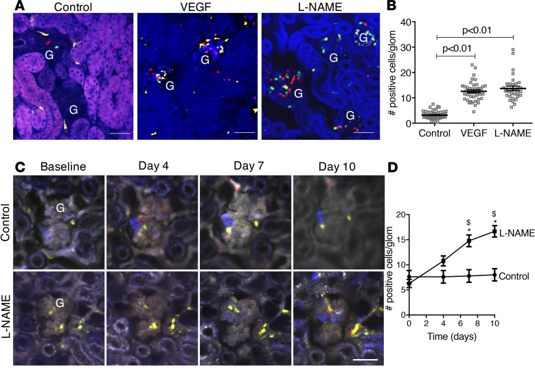
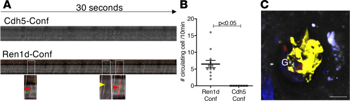
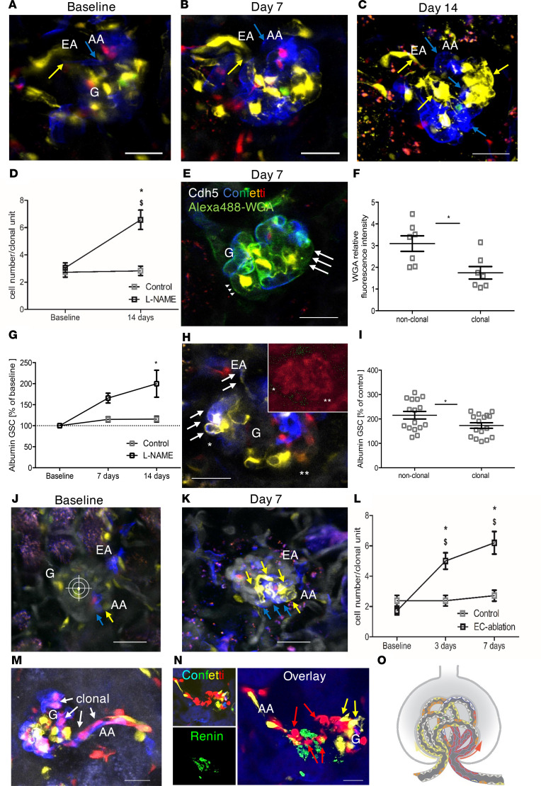
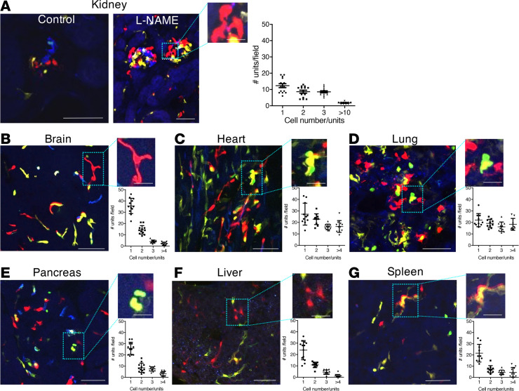
Endothelial cells are important in the maintenance of healthy blood vessels and in the development of vascular diseases. However, the origin and dynamics of endothelial precursors and remodeling at the single-cell level have been difficult to study in vivo owing to technical limitations. Therefore, we aimed to develop a direct visual approach to track the fate and function of single endothelial cells over several days and weeks in the same vascular bed in vivo using multiphoton microscopy (MPM) of transgenic Cdh5-Confetti mice and the kidney glomerulus as a model. Individual cells of the vascular endothelial lineage were identified and tracked owing to their unique color combination, based on the random expression of cyan/green/yellow/red fluorescent proteins. Experimental hypertension, hyperglycemia, and laser-induced endothelial cell ablation rapidly increased the number of new glomerular endothelial cells that appeared in clusters of the same color, suggesting clonal cell remodeling by local precursors at the vascular pole. Furthermore, intravital MPM allowed the detection of distinct structural and functional alterations of proliferating endothelial cells. No circulating Cdh5-Confetti+ cells were found in the renal cortex. Moreover, the heart, lung, and kidneys showed more significant clonal endothelial cell expansion compared with the brain, pancreas, liver, and spleen. In summary, we have demonstrated that serial MPM of Cdh5-Confetti mice in vivo is a powerful technical advance to study endothelial remodeling and repair in the kidney and other organs under physiological and disease conditions.
内皮细胞在维持健康的血管和血管疾病的发展中起着重要作用。然而,由于技术限制,内皮前体细胞的起源和动力学以及单细胞水平的重塑一直难以在体内进行研究。因此,我们旨在开发一种直接的可视化方法,使用多光子显微镜(MPM)对转基因 Cdh5-Confetti 小鼠和肾脏肾小球进行研究,以追踪单个内皮细胞在相同血管床中的命运和功能,时间跨度为数天到数周。由于青色/绿色/黄色/红色荧光蛋白的随机表达,单个血管内皮谱系的细胞可以通过其独特的颜色组合被识别和追踪。实验性高血压、高血糖和激光诱导的内皮细胞消融会迅速增加新的肾小球内皮细胞的数量,这些细胞出现在相同颜色的簇中,这表明血管极处的局部前体细胞通过克隆细胞重塑。此外,活体 MPM 允许检测到增殖内皮细胞的不同结构和功能改变。在肾皮质中未发现循环的 Cdh5-Confetti+细胞。此外,与脑、胰腺、肝和脾相比,心脏、肺和肾脏的克隆内皮细胞扩张更为明显。总之,我们已经证明,体内 Cdh5-Confetti 小鼠的连续 MPM 是研究生理和疾病条件下肾脏和其他器官内皮重塑和修复的强大技术进步。