Villouta Camilo, Cox Benjamin L, Rauch Beth, Workmaster Beth Ann A, Eliceiri Kevin W, Atucha Amaya
Department of Horticulture, University of Wisconsin-Madison, 1575 Linden Dr., Madison, WI, 53706, USA.
Medical Engineering Group, Morgridge Institute for Research, 330 N Orchard St, Madison, WI, 53706, USA.
Plant Methods. 2021 Apr 13;17(1):41. doi: 10.1186/s13007-021-00743-4.
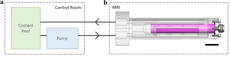
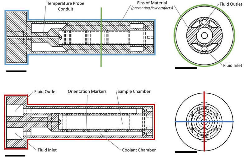
Investigating plant mechanisms to tolerate freezing temperatures is critical to developing crops with superior cold hardiness. However, the lack of imaging methods that allow the visualization of freezing events in complex plant tissues remains a key limitation. Magnetic resonance imaging (MRI) has been successfully used to study many different plant models, including the study of in vivo changes during freezing. However, despite its benefits and past successes, the use of MRI in plant sciences remains low, likely due to limited access, high costs, and associated engineering challenges, such as keeping samples frozen for cold hardiness studies. To address this latter need, a novel device for keeping plant specimens at freezing temperatures during MRI is described.
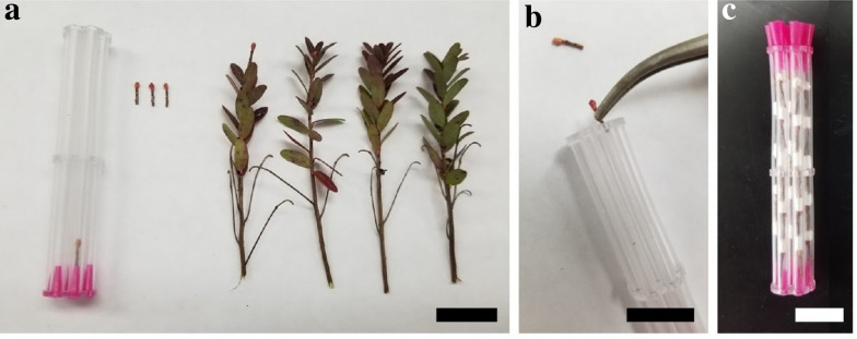
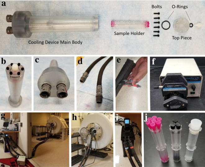
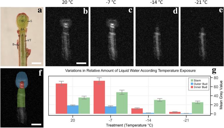
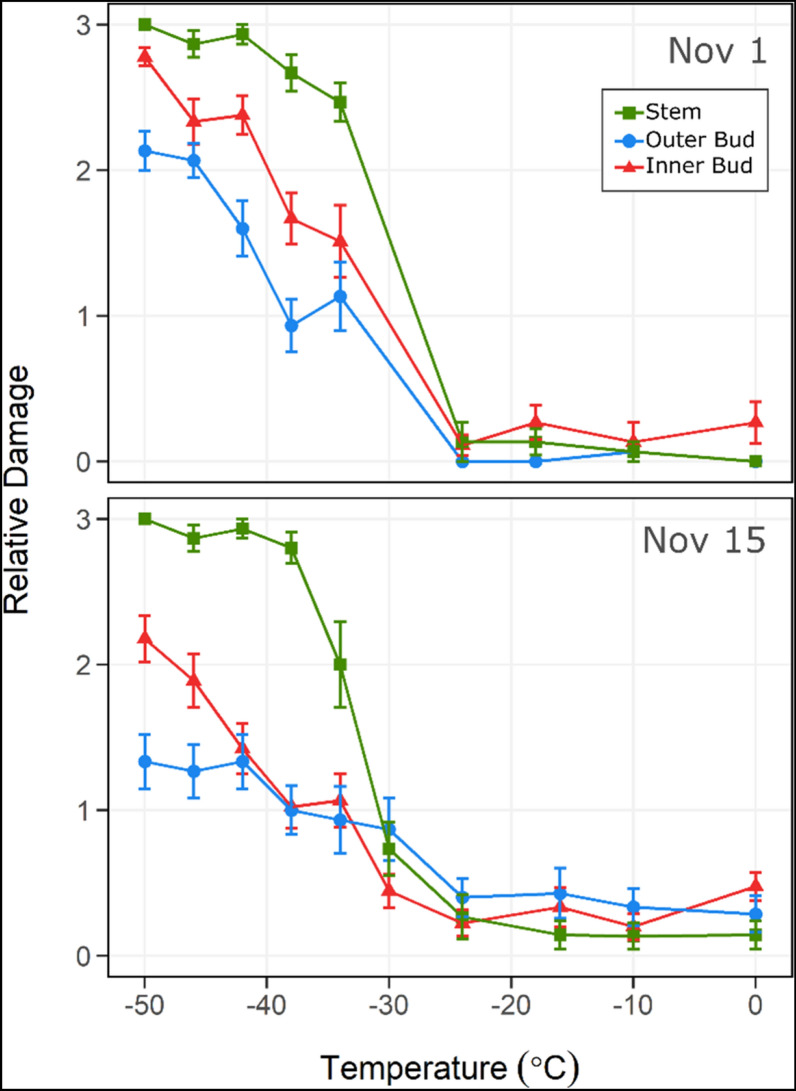
The device consists of commercial and custom parts. All custom parts were 3D printed and made available as open source to increase accessibility to research groups who wish to reproduce or iterate on this work. Calibration tests documented that, upon temperature equilibration for a given experimental temperature, conditions between the circulating coolant bath and inside the device seated within the bore of the magnet varied by less than 0.1 °C. The device was tested on plant material by imaging buds from Vaccinium macrocarpon in a small animal MRI system, at four temperatures, 20 °C, - 7 °C, - 14 °C, and - 21 °C. Results were compared to those obtained by independent controlled freezing test (CFT) evaluations. Non-damaging freezing events in inner bud structures were detected from the imaging data collected using this device, phenomena that are undetectable using CFT.
The use of this novel cooling and freezing device in conjunction with MRI facilitated the detection of freezing events in intact plant tissues through the observation of the presence and absence of water in liquid state. The device represents an important addition to plant imaging tools currently available to researchers. Furthermore, its open-source and customizable design ensures that it will be accessible to a wide range of researchers and applications.
研究植物耐受低温的机制对于培育具有卓越抗寒能力的作物至关重要。然而,缺乏能够可视化复杂植物组织中冷冻事件的成像方法仍然是一个关键限制。磁共振成像(MRI)已成功用于研究许多不同的植物模型,包括冷冻过程中的体内变化研究。然而,尽管MRI有诸多优点且过去取得了成功,但它在植物科学中的应用仍然较少,这可能是由于设备使用受限、成本高昂以及相关的工程挑战,例如在进行抗寒研究时要使样本保持冷冻状态。为满足这一需求,本文描述了一种在MRI过程中使植物标本保持冷冻温度的新型装置。
该装置由商业部件和定制部件组成。所有定制部件均通过3D打印制作,并作为开源提供,以方便希望重复或改进此项工作的研究团队使用。校准测试表明,在给定实验温度下达到温度平衡后,循环冷却浴与置于磁体孔内的装置内部之间的温度差异小于0.1°C。该装置在小型动物MRI系统中对四种温度(20°C、-7°C、-14°C和-21°C)下的大果越桔芽进行成像测试。结果与通过独立控制冷冻试验(CFT)评估获得的结果进行了比较。从使用该装置收集的成像数据中检测到了内部芽结构中的非损伤性冷冻事件,而这些现象是使用CFT无法检测到的。
结合MRI使用这种新型冷却和冷冻装置,通过观察液态水的有无,有助于检测完整植物组织中的冷冻事件。该装置是研究人员目前可用的植物成像工具的重要补充。此外,其开源和可定制设计确保了广泛的研究人员能够使用并应用于多种研究。