• 文献检索
  • 文档翻译
  • 深度研究
  • 学术资讯
  • Suppr Zotero 插件Zotero 插件
  • 邀请有礼
  • 套餐&价格
  • 历史记录
应用&插件
Suppr Zotero 插件Zotero 插件浏览器插件Mac 客户端Windows 客户端微信小程序
定价
高级版会员购买积分包购买API积分包
服务
文献检索文档翻译深度研究API 文档MCP 服务
关于我们
关于 Suppr公司介绍联系我们用户协议隐私条款
关注我们

Suppr 超能文献

核心技术专利:CN118964589B侵权必究
粤ICP备2023148730 号-1Suppr @ 2026

文献检索

告别复杂PubMed语法,用中文像聊天一样搜索,搜遍4000万医学文献。AI智能推荐,让科研检索更轻松。

立即免费搜索

文件翻译

保留排版,准确专业,支持PDF/Word/PPT等文件格式,支持 12+语言互译。

免费翻译文档

深度研究

AI帮你快速写综述,25分钟生成高质量综述,智能提取关键信息,辅助科研写作。

立即免费体验

基于胸部 X 光的人工智能肺炎诊断模型的性能。

Diagnostic performance of artificial intelligence model for pneumonia from chest radiography.

机构信息

JLK, Incorporated, Eonju-ro, Gangnam-gu, Seoul, South Korea.

Department of Internal Medicine, Gil Medical Center, Gachon University College of Medicine, Incheon, South Korea.

出版信息

PLoS One. 2021 Apr 15;16(4):e0249399. doi: 10.1371/journal.pone.0249399. eCollection 2021.

DOI:10.1371/journal.pone.0249399
PMID:33857181
原文链接:https://pmc.ncbi.nlm.nih.gov/articles/PMC8049482/
Abstract

OBJECTIVE

The chest X-ray (CXR) is the most readily available and common imaging modality for the assessment of pneumonia. However, detecting pneumonia from chest radiography is a challenging task, even for experienced radiologists. An artificial intelligence (AI) model might help to diagnose pneumonia from CXR more quickly and accurately. We aim to develop an AI model for pneumonia from CXR images and to evaluate diagnostic performance with external dataset.

METHODS

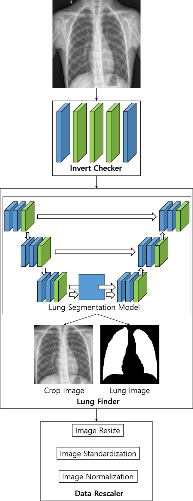
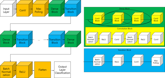
To train the pneumonia model, a total of 157,016 CXR images from the National Institutes of Health (NIH) and the Korean National Tuberculosis Association (KNTA) were used (normal vs. pneumonia = 120,722 vs.36,294). An ensemble model of two neural networks with DenseNet classifies each CXR image into pneumonia or not. To test the accuracy of the models, a separate external dataset of pneumonia CXR images (n = 212) from a tertiary university hospital (Gachon University Gil Medical Center GUGMC, Incheon, South Korea) was used; the diagnosis of pneumonia was based on both the chest CT findings and clinical information, and the performance evaluated using the area under the receiver operating characteristic curve (AUC). Moreover, we tested the change of the AI probability score for pneumonia using the follow-up CXR images (7 days after the diagnosis of pneumonia, n = 100).

RESULTS

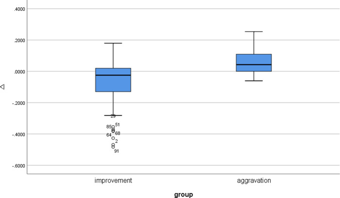
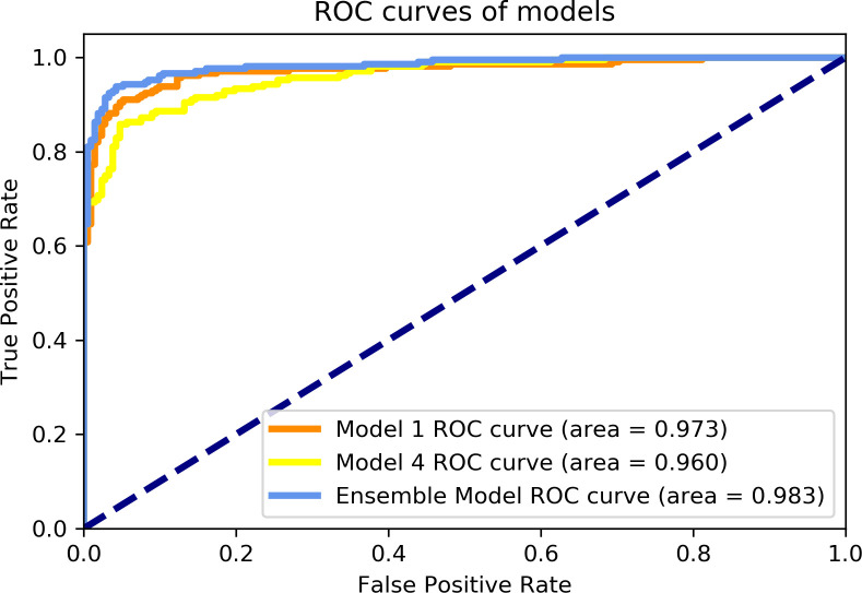
When the probability scores of the models that have a threshold of 0.5 for pneumonia, two models (models 1 and 4) having different pre-processing parameters on the histogram equalization distribution showed best AUC performances of 0.973 and 0.960, respectively. As expected, the ensemble model of these two models performed better than each of the classification models with 0.983 AUC. Furthermore, the AI probability score change for pneumonia showed a significant difference between improved cases and aggravated cases (Δ = -0.06 ± 0.14 vs. 0.06 ± 0.09, for 85 improved cases and 15 aggravated cases, respectively, P = 0.001) for CXR taken as a 7-day follow-up.

CONCLUSIONS

The ensemble model combined two different classification models for pneumonia that performed at 0.983 AUC for an external test dataset from a completely different data source. Furthermore, AI probability scores showed significant changes between cases of different clinical prognosis, which suggest the possibility of increased efficiency and performance of the CXR reading at the diagnosis and follow-up evaluation for pneumonia.

摘要

目的

胸部 X 光(CXR)是评估肺炎最常用的成像方式。然而,即使对于有经验的放射科医生来说,从胸部 X 光片中检测肺炎也是一项具有挑战性的任务。人工智能(AI)模型可能有助于更快、更准确地从 CXR 诊断肺炎。我们旨在开发一种用于 CXR 图像的肺炎 AI 模型,并使用外部数据集评估诊断性能。

方法

为了训练肺炎模型,共使用了来自美国国立卫生研究院(NIH)和韩国国家结核病协会(KNTA)的 157016 张 CXR 图像(正常 vs. 肺炎=120722 比 36294)。两个带有 DenseNet 的神经网络的集成模型将每张 CXR 图像分为肺炎或非肺炎。为了测试模型的准确性,使用了一家三级大学医院(韩国仁川加图大学吉尔医疗中心 GUGMC)的另一个外部肺炎 CXR 图像数据集(n=212);肺炎的诊断基于胸部 CT 结果和临床信息,并使用接收者操作特征曲线下的面积(AUC)进行评估。此外,我们还测试了使用后续 CXR 图像(肺炎诊断后 7 天,n=100)时 AI 肺炎概率评分的变化。

结果

当概率评分模型的阈值为 0.5 时,两个具有不同直方图均衡分布预处理参数的模型(模型 1 和 4)表现出最佳的 AUC 性能,分别为 0.973 和 0.960。如预期的那样,这两个模型的集成模型表现优于每个分类模型,AUC 为 0.983。此外,肺炎的 AI 概率评分变化在改善病例和加重病例之间有显著差异(Δ=-0.06±0.14 比 0.06±0.09,分别为 85 例改善病例和 15 例加重病例,P=0.001),这些病例是在 CXR 拍摄的 7 天随访时的结果。

结论

该集成模型结合了两种不同的肺炎分类模型,在完全不同数据源的外部测试数据集上的 AUC 为 0.983。此外,AI 概率评分在不同临床预后病例之间有显著变化,这表明 CXR 阅读在肺炎的诊断和随访评估中的效率和性能可能会提高。

https://cdn.ncbi.nlm.nih.gov/pmc/blobs/9964/8049482/f77de5caf85f/pone.0249399.g006.jpg
https://cdn.ncbi.nlm.nih.gov/pmc/blobs/9964/8049482/664bfc2c06d8/pone.0249399.g001.jpg
https://cdn.ncbi.nlm.nih.gov/pmc/blobs/9964/8049482/410d49fb080f/pone.0249399.g002.jpg
https://cdn.ncbi.nlm.nih.gov/pmc/blobs/9964/8049482/90137d9d1d4d/pone.0249399.g003.jpg
https://cdn.ncbi.nlm.nih.gov/pmc/blobs/9964/8049482/d8e27b9333e5/pone.0249399.g004.jpg
https://cdn.ncbi.nlm.nih.gov/pmc/blobs/9964/8049482/22ef1ba79048/pone.0249399.g005.jpg
https://cdn.ncbi.nlm.nih.gov/pmc/blobs/9964/8049482/f77de5caf85f/pone.0249399.g006.jpg

相似文献

1
Diagnostic performance of artificial intelligence model for pneumonia from chest radiography.基于胸部 X 光的人工智能肺炎诊断模型的性能。
PLoS One. 2021 Apr 15;16(4):e0249399. doi: 10.1371/journal.pone.0249399. eCollection 2021.
2
Thorax computed tomography (CTX) guided ground truth annotation of CHEST radiographs (CXR) for improved classification and detection of COVID-19.胸部 CT 引导的 CHEST 射线照片(CXR)标注以提高 COVID-19 的分类和检测。
Int J Numer Method Biomed Eng. 2024 Jun;40(6):e3823. doi: 10.1002/cnm.3823. Epub 2024 Apr 8.
3
AI-based computer-aided diagnostic system of chest digital tomography synthesis: Demonstrating comparative advantage with X-ray-based AI systems.基于人工智能的胸部数字断层合成计算机辅助诊断系统:与基于 X 射线的人工智能系统比较优势展示。
Comput Methods Programs Biomed. 2023 Oct;240:107643. doi: 10.1016/j.cmpb.2023.107643. Epub 2023 Jun 5.
4
Multi-View Ensemble Convolutional Neural Network to Improve Classification of Pneumonia in Low Contrast Chest X-Ray Images.多视图集成卷积神经网络用于改善低对比度胸部X光图像中肺炎的分类
Annu Int Conf IEEE Eng Med Biol Soc. 2020 Jul;2020:1238-1241. doi: 10.1109/EMBC44109.2020.9176517.
5
Comparison of Chest Radiograph Interpretations by Artificial Intelligence Algorithm vs Radiology Residents.人工智能算法与放射科住院医师对胸部 X 线片解读的比较。
JAMA Netw Open. 2020 Oct 1;3(10):e2022779. doi: 10.1001/jamanetworkopen.2020.22779.
6
Artificial Intelligence Algorithm Detecting Lung Infection in Supine Chest Radiographs of Critically Ill Patients With a Diagnostic Accuracy Similar to Board-Certified Radiologists.人工智能算法检测重症患者仰卧位胸部 X 光片肺部感染的诊断准确性与放射科认证医师相当。
Crit Care Med. 2020 Jul;48(7):e574-e583. doi: 10.1097/CCM.0000000000004397.
7
Tuberculosis detection from chest x-rays for triaging in a high tuberculosis-burden setting: an evaluation of five artificial intelligence algorithms.从高结核病负担环境中的胸部 X 光片中检测结核病以进行分诊:五种人工智能算法的评估。
Lancet Digit Health. 2021 Sep;3(9):e543-e554. doi: 10.1016/S2589-7500(21)00116-3.
8
Association of Artificial Intelligence-Aided Chest Radiograph Interpretation With Reader Performance and Efficiency.人工智能辅助的胸部 X 光片解读与读者表现和效率的关联。
JAMA Netw Open. 2022 Aug 1;5(8):e2229289. doi: 10.1001/jamanetworkopen.2022.29289.
9
Using deep-learning techniques for pulmonary-thoracic segmentations and improvement of pneumonia diagnosis in pediatric chest radiographs.使用深度学习技术进行肺胸段分割,提高儿科胸部 X 光片中肺炎的诊断准确率。
Pediatr Pulmonol. 2019 Oct;54(10):1617-1626. doi: 10.1002/ppul.24431. Epub 2019 Jul 3.
10
Deep Learning Models to Predict Fatal Pneumonia Using Chest X-Ray Images.深度学习模型利用胸部 X 光图像预测致命性肺炎。
Can Respir J. 2022 Nov 24;2022:8026580. doi: 10.1155/2022/8026580. eCollection 2022.

引用本文的文献

1
AI for Detection of Tuberculosis: Implications for Global Health.人工智能在结核病检测中的应用:对全球健康的影响。
Radiol Artif Intell. 2024 Mar;6(2):e230327. doi: 10.1148/ryai.230327.
2
Incidence and Outcomes of Non-Ventilator-Associated Hospital-Acquired Pneumonia in 284 US Hospitals Using Electronic Surveillance Criteria.电子监测标准下 284 家美国医院中非呼吸机相关性医院获得性肺炎的发生率和结局。
JAMA Netw Open. 2023 May 1;6(5):e2314185. doi: 10.1001/jamanetworkopen.2023.14185.
3
Incidentally found resectable lung cancer with the usage of artificial intelligence on chest radiographs.

本文引用的文献

1
Artificial intelligence applications for thoracic imaging.人工智能在胸部成像中的应用。
Eur J Radiol. 2020 Feb;123:108774. doi: 10.1016/j.ejrad.2019.108774. Epub 2019 Dec 11.
2
Deep Learning for Chest Radiograph Diagnosis in the Emergency Department.深度学习在急诊科胸部 X 光诊断中的应用。
Radiology. 2019 Dec;293(3):573-580. doi: 10.1148/radiol.2019191225. Epub 2019 Oct 22.
3
Development and Validation of a Deep Learning-Based Automated Detection Algorithm for Major Thoracic Diseases on Chest Radiographs.基于深度学习的胸部 X 线片主要胸部疾病自动检测算法的开发与验证。
在胸部 X 光片使用人工智能时偶然发现可切除的肺癌。
PLoS One. 2023 Mar 10;18(3):e0281690. doi: 10.1371/journal.pone.0281690. eCollection 2023.
4
Chest X-ray in Emergency Radiology: What Artificial Intelligence Applications Are Available?急诊放射学中的胸部X光检查:有哪些可用的人工智能应用?
Diagnostics (Basel). 2023 Jan 6;13(2):216. doi: 10.3390/diagnostics13020216.
5
Deep Learning in Multi-Class Lung Diseases' Classification on Chest X-ray Images.深度学习在胸部X光图像多类肺部疾病分类中的应用
Diagnostics (Basel). 2022 Apr 6;12(4):915. doi: 10.3390/diagnostics12040915.
JAMA Netw Open. 2019 Mar 1;2(3):e191095. doi: 10.1001/jamanetworkopen.2019.1095.
4
Deep learning for chest radiograph diagnosis: A retrospective comparison of the CheXNeXt algorithm to practicing radiologists.深度学习在胸片诊断中的应用:CheXNeXt 算法与临床放射科医生的回顾性比较。
PLoS Med. 2018 Nov 20;15(11):e1002686. doi: 10.1371/journal.pmed.1002686. eCollection 2018 Nov.
5
Development and Validation of a Deep Learning-based Automatic Detection Algorithm for Active Pulmonary Tuberculosis on Chest Radiographs.基于深度学习的胸部 X 线片活动性肺结核自动检测算法的开发与验证。
Clin Infect Dis. 2019 Aug 16;69(5):739-747. doi: 10.1093/cid/ciy967.
6
Development and Validation of Deep Learning-based Automatic Detection Algorithm for Malignant Pulmonary Nodules on Chest Radiographs.基于深度学习的胸部 X 线片恶性肺结节自动检测算法的开发与验证。
Radiology. 2019 Jan;290(1):218-228. doi: 10.1148/radiol.2018180237. Epub 2018 Sep 25.
7
Computer-aided detection in chest radiography based on artificial intelligence: a survey.基于人工智能的胸部 X 射线计算机辅助检测:综述。
Biomed Eng Online. 2018 Aug 22;17(1):113. doi: 10.1186/s12938-018-0544-y.
8
Preliminary report from the World Health Organisation Chest Radiography in Epidemiological Studies project.世界卫生组织“流行病学研究中的胸部X光摄影”项目的初步报告。
Pediatr Radiol. 2017 Oct;47(11):1399-1404. doi: 10.1007/s00247-017-3834-9. Epub 2017 Sep 21.
9
Viruses and bacteria in sputum samples of children with community-acquired pneumonia.痰液样本中的病毒和细菌与社区获得性肺炎的儿童。
Clin Microbiol Infect. 2012 Mar;18(3):300-7. doi: 10.1111/j.1469-0691.2011.03603.x. Epub 2011 Aug 18.
10
Viral pneumonia.病毒性肺炎。
Lancet. 2011 Apr 9;377(9773):1264-75. doi: 10.1016/S0140-6736(10)61459-6. Epub 2011 Mar 22.