• 文献检索
  • 文档翻译
  • 深度研究
  • 学术资讯
  • Suppr Zotero 插件Zotero 插件
  • 邀请有礼
  • 套餐&价格
  • 历史记录
应用&插件
Suppr Zotero 插件Zotero 插件浏览器插件Mac 客户端Windows 客户端微信小程序
定价
高级版会员购买积分包购买API积分包
服务
文献检索文档翻译深度研究API 文档MCP 服务
关于我们
关于 Suppr公司介绍联系我们用户协议隐私条款
关注我们

Suppr 超能文献

核心技术专利:CN118964589B侵权必究
粤ICP备2023148730 号-1Suppr @ 2026

文献检索

告别复杂PubMed语法,用中文像聊天一样搜索,搜遍4000万医学文献。AI智能推荐,让科研检索更轻松。

立即免费搜索

文件翻译

保留排版,准确专业,支持PDF/Word/PPT等文件格式,支持 12+语言互译。

免费翻译文档

深度研究

AI帮你快速写综述,25分钟生成高质量综述,智能提取关键信息,辅助科研写作。

立即免费体验

术中光学相干断层扫描引导角膜清扫术在超薄Descemet膜剥除自动内皮角膜移植术中用于清除残余界面液。

Intraoperative optical coherence tomography guided corneal sweeping for removal of remnant Interface fluid during ultra-thin Descemet stripping automated endothelial keratoplasty.

作者信息

Mimouni Michael, Kronschläger Martin, Ruiss Manuel, Findl Oliver

机构信息

Department of Ophthalmology, Rambam Health Care Campus, Haifa affiliated with the Bruce and Ruth Rappaport Faculty of Medicine, Technion-Israel Institute of Technology, Haifa, Israel.

Department of Ophthalmology and Vision Sciences, University of Toronto, Toronto, Ontario, Canada.

出版信息

BMC Ophthalmol. 2021 Apr 15;21(1):180. doi: 10.1186/s12886-021-01934-2.

DOI:10.1186/s12886-021-01934-2
PMID:33858385
原文链接:https://pmc.ncbi.nlm.nih.gov/articles/PMC8048159/
Abstract

BACKGROUND

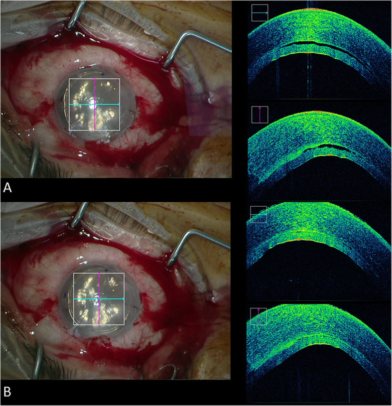
Remnant interface fluid following Descemet stripping automated endothelial keratoplasty (DSAEK) is associated with postoperative detachments. The aim of this study was to assess outcomes of intraoperative optical coherence tomography (iOCT) guided meticulous peripheral corneal sweeping for removal of interface fluid during ultra-thin (UT) DSAEK.

METHODS

This retrospective study included all eyes underwent iOCT guided UT-DSAEK from October 2016 to February 2018 at the Hanusch Hospital, Vienna, Austria. Peripheral meticulous corneal sweeping was performed to remove excess fluid. Central graft thickness (CGT) was measured prior to surgery, after graft bubbling and after corneal sweeping. Remnant interface fluid rates were compared between eyes that underwent rebubbling and those that did not.

RESULTS

Overall, 28 eyes of 28 patients with a mean age of 73.9 ± 10.0 years were included. An iOCT guided meticulous peripheral sweeping was performed in 89.3% (n = 25) of the cases. Following 84% (n = 21) of the peripheral sweeping performed, remnant fluid was no longer identified. Following peripheral sweeping the interface fluid height was reduced from 17.31 ± 15.96 μm to 3.46 ± 9.52 μm (p < 0.001) and CGT was reduced by 7% (p < 0.001). Rebubbling was performed in 17.9% (n = 5) of the cases. The rebubbling group had a greater proportion of patients that had remnant fluid identified with iOCT at the end of surgery despite meticulous peripheral sweeping (60.0% versus 4.4%, p = 0.01).

CONCLUSION

The iOCT identified subclinical remnant fluid in nearly 90% of UT-DSAEK cases. An iOCT guided peripheral corneal sweeping led to resolution of interface fluid in a majority of cases. Eyes with persistent remnant fluid despite peripheral corneal sweeping are more likely to require subsequent rebubbling.

摘要

背景

在Descemet膜剥除自动内皮角膜移植术(DSAEK)后,残留的界面液体会导致术后角膜瓣脱离。本研究的目的是评估术中光学相干断层扫描(iOCT)引导下在超薄(UT)DSAEK手术中进行细致的周边角膜清扫以去除界面液体的效果。

方法

这项回顾性研究纳入了2016年10月至2018年2月在奥地利维也纳哈努施医院接受iOCT引导的UT-DSAEK手术的所有患眼。进行周边细致的角膜清扫以去除多余的液体。在手术前、植片形成水泡后和角膜清扫后测量中央植片厚度(CGT)。比较进行再次气泡形成的患眼和未进行再次气泡形成的患眼之间的残留界面液率。

结果

总体而言,纳入了28例患者的28只患眼,平均年龄为73.9±10.0岁。89.3%(n = 25)的病例进行了iOCT引导下的周边细致清扫。在84%(n = 21)的周边清扫后,未再发现残留液体。周边清扫后,界面液体高度从17.31±15.96μm降至至3.46±9.52μm(p < 0.001),CGT降低了7%(p < 0.001)。17.9%(n = 5)的病例进行了再次气泡形成。再次气泡形成组中,尽管进行了细致的周边清扫,但在手术结束时通过iOCT发现仍有残留液体的患者比例更高(60.0%对4.4%,p = 0.01)。

结论

iOCT在近90%的UT-DSAEK病例中发现了亚临床残留液体。iOCT引导下的周边角膜清扫在大多数病例中可使界面液体得到解决。尽管进行了周边角膜清扫但仍有持续残留液体的患眼更有可能需要随后的再次气泡形成。

https://cdn.ncbi.nlm.nih.gov/pmc/blobs/a99e/8048159/260dbf5e33ea/12886_2021_1934_Fig3_HTML.jpg
https://cdn.ncbi.nlm.nih.gov/pmc/blobs/a99e/8048159/61fdebf6b80c/12886_2021_1934_Fig1_HTML.jpg
https://cdn.ncbi.nlm.nih.gov/pmc/blobs/a99e/8048159/e66492997759/12886_2021_1934_Fig2_HTML.jpg
https://cdn.ncbi.nlm.nih.gov/pmc/blobs/a99e/8048159/260dbf5e33ea/12886_2021_1934_Fig3_HTML.jpg
https://cdn.ncbi.nlm.nih.gov/pmc/blobs/a99e/8048159/61fdebf6b80c/12886_2021_1934_Fig1_HTML.jpg
https://cdn.ncbi.nlm.nih.gov/pmc/blobs/a99e/8048159/e66492997759/12886_2021_1934_Fig2_HTML.jpg
https://cdn.ncbi.nlm.nih.gov/pmc/blobs/a99e/8048159/260dbf5e33ea/12886_2021_1934_Fig3_HTML.jpg

相似文献

1
Intraoperative optical coherence tomography guided corneal sweeping for removal of remnant Interface fluid during ultra-thin Descemet stripping automated endothelial keratoplasty.术中光学相干断层扫描引导角膜清扫术在超薄Descemet膜剥除自动内皮角膜移植术中用于清除残余界面液。
BMC Ophthalmol. 2021 Apr 15;21(1):180. doi: 10.1186/s12886-021-01934-2.
2
Intraoperative Interface Fluid Dynamics and Clinical Outcomes for Intraoperative Optical Coherence Tomography-Assisted Descemet Stripping Automated Endothelial Keratoplasty From the PIONEER Study.先锋研究中术中光学相干断层扫描辅助的后弹力层剥除自动角膜内皮移植术的术中界面流体动力学及临床结果
Am J Ophthalmol. 2017 Jan;173:16-22. doi: 10.1016/j.ajo.2016.09.028. Epub 2016 Oct 1.
3
Association between transient interface fluid on intraoperative OCT and textural interface opacity after DSAEK surgery in the PIONEER study.PIONEER 研究中术中 OCT 上的瞬态界面液与 DSAEK 手术后的纹理状界面不透明之间的关联。
Cornea. 2014 Sep;33(9):887-92. doi: 10.1097/ICO.0000000000000209.
4
Automated volumetric analysis of interface fluid in descemet stripping automated endothelial keratoplasty using intraoperative optical coherence tomography.使用术中光相干断层扫描对撕囊自动角膜内皮移植术中界面液进行自动容积分析。
Invest Ophthalmol Vis Sci. 2014 Aug 7;55(9):5610-5. doi: 10.1167/iovs.14-14346.
5
Intraoperative optical coherence tomography in descemet stripping automated endothelial keratoplasty: pilot experiences.Descemet膜剥除自动内皮角膜移植术中的术中光学相干断层扫描:初步经验
Int Ophthalmol. 2017 Aug;37(4):939-944. doi: 10.1007/s10792-016-0338-9. Epub 2016 Sep 21.
6
Evaluation of Time to Donor Lenticule Apposition Using Intraoperative Optical Coherence Tomography in Descemet Stripping Automated Endothelial Keratoplasty.在深板层角膜内皮移植术中使用术中光学相干断层扫描评估供体角膜瓣贴合时间
Cornea. 2016 Apr;35(4):477-81. doi: 10.1097/ICO.0000000000000757.
7
Anterior Segment Optical Coherence Tomography of Post-Descemet Stripping Automated Endothelial Keratoplasty Eyes to Evaluate Graft Morphology and Its Association With Visual Outcome.后弹力层剥除自动角膜内皮移植术后眼睛的眼前节光学相干断层扫描,以评估移植物形态及其与视觉结果的关联。
Cornea. 2018 Sep;37(9):1087-1092. doi: 10.1097/ICO.0000000000001596.
8
Complications of Descemet's stripping with automated endothelial keratoplasty: survey of 118 eyes at One Institute.自动角膜内皮移植术行后弹力层剥除的并发症:一所机构118只眼的调查
Ophthalmology. 2008 Sep;115(9):1517-24. doi: 10.1016/j.ophtha.2008.01.024. Epub 2008 Apr 18.
9
Intraoperative Optical Coherence Tomography-Assisted Descemet Membrane Endothelial Keratoplasty in the DISCOVER Study: First 100 Cases.术中光学相干断层扫描辅助 Descemet 膜内皮角膜移植术在 DISCOVER 研究中的应用:前 100 例。
Am J Ophthalmol. 2020 Feb;210:167-173. doi: 10.1016/j.ajo.2019.09.018. Epub 2019 Sep 25.
10
Effects of graft thickness and asymmetry on visual gain and aberrations after descemet stripping automated endothelial keratoplasty.移植片厚度和不对称性对撕囊自动化角膜内皮移植术后视觉增益和像差的影响。
JAMA Ophthalmol. 2013 Jun;131(6):737-44. doi: 10.1001/jamaophthalmol.2013.73.

引用本文的文献

1
Lomi Lomi massage for clearing interface fluid in Descemet stripping automated endothelial keratoplasty.用于在Descemet膜剥离自动内皮角膜移植术中清除界面液体的洛米洛米按摩法
Indian J Ophthalmol. 2025 Jul 1;73(7):1088. doi: 10.4103/IJO.IJO_2957_24. Epub 2025 Jun 30.
2
Expanding Application of Optical Coherence Tomography Beyond the Clinic: A Narrative Review.光学相干断层扫描在临床之外的扩展应用:一项叙述性综述
Diagnostics (Basel). 2025 Apr 29;15(9):1140. doi: 10.3390/diagnostics15091140.

本文引用的文献

1
Intraoperative Optical Coherence Tomography-Assisted Descemet Membrane Endothelial Keratoplasty in the DISCOVER Study: First 100 Cases.术中光学相干断层扫描辅助 Descemet 膜内皮角膜移植术在 DISCOVER 研究中的应用:前 100 例。
Am J Ophthalmol. 2020 Feb;210:167-173. doi: 10.1016/j.ajo.2019.09.018. Epub 2019 Sep 25.
2
Immune reactions after modern lamellar (DALK, DSAEK, DMEK) versus conventional penetrating corneal transplantation.现代板层角膜移植术(DALK、DSAEK、DMEK)与传统穿透性角膜移植术后的免疫反应。
Prog Retin Eye Res. 2019 Nov;73:100768. doi: 10.1016/j.preteyeres.2019.07.001. Epub 2019 Jul 3.
3
New trends in corneal transplants at the University of Toronto.
多伦多大学角膜移植的新趋势。
Can J Ophthalmol. 2018 Dec;53(6):580-587. doi: 10.1016/j.jcjo.2018.02.023. Epub 2018 May 9.
4
Intraoperative Optical Coherence Tomography-Guided Membrane Peeling for Surgery of Macular Pucker: Advantages and Limitations.术中光学相干断层扫描引导下的黄斑皱襞手术剥膜:优势与局限性
Ophthalmologica. 2019;241(4):234-240. doi: 10.1159/000493279. Epub 2018 Nov 9.
5
Intraoperative optical coherence tomography measurements of aphakic eyes to predict postoperative position of 2 intraocular lens designs.术中光学相干断层扫描测量术对预测两种人工晶状体设计术后位置的研究。
J Cataract Refract Surg. 2018 Nov;44(11):1310-1316. doi: 10.1016/j.jcrs.2018.07.044. Epub 2018 Sep 12.
6
Trends in Corneal Transplantation from 2001 to 2016 in Germany: A Report of the DOG-Section Cornea and its Keratoplasty Registry.2001 年至 2016 年德国角膜移植趋势:德国眼库协会角膜及其移植登记报告。
Am J Ophthalmol. 2018 Apr;188:91-98. doi: 10.1016/j.ajo.2018.01.018. Epub 2018 Feb 2.
7
Factors Associated With Graft Detachment After Primary Descemet Stripping Automated Endothelial Keratoplasty.原发性后弹力层剥除自动角膜内皮移植术后移植物脱离的相关因素
Cornea. 2017 Mar;36(3):265-268. doi: 10.1097/ICO.0000000000001123.
8
Intraoperative Interface Fluid Dynamics and Clinical Outcomes for Intraoperative Optical Coherence Tomography-Assisted Descemet Stripping Automated Endothelial Keratoplasty From the PIONEER Study.先锋研究中术中光学相干断层扫描辅助的后弹力层剥除自动角膜内皮移植术的术中界面流体动力学及临床结果
Am J Ophthalmol. 2017 Jan;173:16-22. doi: 10.1016/j.ajo.2016.09.028. Epub 2016 Oct 1.
9
Intraoperative optical coherence tomography in descemet stripping automated endothelial keratoplasty: pilot experiences.Descemet膜剥除自动内皮角膜移植术中的术中光学相干断层扫描:初步经验
Int Ophthalmol. 2017 Aug;37(4):939-944. doi: 10.1007/s10792-016-0338-9. Epub 2016 Sep 21.
10
Visualizing Macular Structures During Membrane Peeling Surgery With an Intraoperative Spectral-Domain Optical Coherence Tomography Device.使用术中光谱域光学相干断层扫描设备在膜剥除手术中可视化黄斑结构
Ophthalmic Surg Lasers Imaging Retina. 2016 Apr 1;47(4):328-32. doi: 10.3928/23258160-20160324-04.