Watanabe Shotaro, Akagi Ryuichiro, Shiko Yuki, Ono Yoshimasa, Kawasaki Yohei, Ohdera Toshihiro, Ohtori Seiji, Sasho Takahisa
Department of Orthopaedic Surgery, Graduate School of Medicine, Development of Orthopaedic Surgery, Chiba University, 1-8-1 Inohana, Chuo-ku, Chiba, Japan.
Center for Preventive Medical Sciences, Chiba University, 1-8-1 Inohana, Chuo-ku, Chiba, Japan.
BMC Musculoskelet Disord. 2021 Apr 17;22(1):363. doi: 10.1186/s12891-021-04228-2.
The evaluation of postoperative total knee arthroplasty (TKA) alignment mainly relies on measurement data obtained from plain radiographs. The aim of this retrospective observational study was to document the intra- and inter-observer reliability in assessment of TKA component positioning after surgery using a three-dimensional (3D) computed tomography (CT) image matching system.
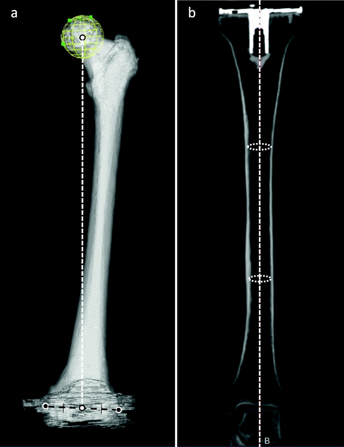
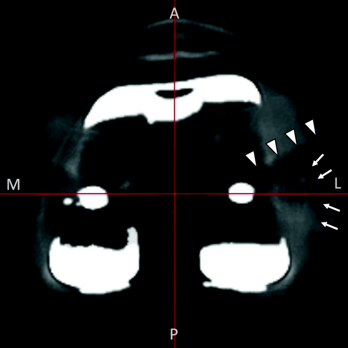
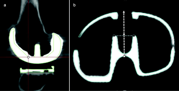
Fourteen knees from 14 patients who received primary TKA were included, and images were analyzed by blinded readers not associated with the surgeries. The examiner digitized the reference points according to defined landmarks, and the designated size component was superimposed to the 3D reconstructed CT model for measurement. In addition to the evaluation of implant position against the coronal and sagittal lower limb mechanical axes that were defined based on bony landmarks, implant position against axes connecting implant-based reference points that are easier to indicate was evaluated.
The overall intra- and inter-observer reliabilities determined by the intraclass correlation coefficients (ICC) of the implant alignment measurement for both femoral and tibial components were good (ICC > 0.60), except in the direction of femoral flexion and extension, for both mechanical and implant-based axes. The difference between implant alignment measurements according to the traditional mechanical axis and the implant-based axis ranged between means of 0.08 and 1.70 and were statistically significantly different.
The postoperative evaluation of implant position in the coronal and sagittal planes using 3D-CT image matching is reliable and has good reproducibility except for the sagittal alignment assessment of the femoral component. The measured implant position according to the traditional mechanical axis and the implant-based axis were slightly but significantly different.
全膝关节置换术(TKA)术后对线评估主要依赖于X线平片获得的测量数据。本回顾性观察研究的目的是使用三维(3D)计算机断层扫描(CT)图像匹配系统记录术后TKA组件定位评估中的观察者内和观察者间可靠性。
纳入14例接受初次TKA患者的14个膝关节,由与手术无关的盲法读者分析图像。检查者根据定义的标志点将参考点数字化,并将指定尺寸的组件叠加到3D重建的CT模型上进行测量。除了根据基于骨性标志定义的冠状面和矢状面下肢机械轴评估植入物位置外,还评估了植入物相对于连接基于植入物的参考点的轴的位置,这些参考点更易于指示。
通过组内相关系数(ICC)确定的股骨和胫骨组件植入物对线测量的总体观察者内和观察者间可靠性良好(ICC>0.60),但在股骨屈伸方向上,对于机械轴和基于植入物的轴均如此。根据传统机械轴和基于植入物的轴进行的植入物对线测量之间的差异在平均值0.08至1.70之间,且具有统计学显著差异。
使用3D-CT图像匹配对冠状面和矢状面植入物位置进行术后评估是可靠的,并且具有良好的可重复性,但股骨组件的矢状面对线评估除外。根据传统机械轴和基于植入物的轴测量的植入物位置略有不同但具有显著差异。