• 文献检索
  • 文档翻译
  • 深度研究
  • 学术资讯
  • Suppr Zotero 插件Zotero 插件
  • 邀请有礼
  • 套餐&价格
  • 历史记录
应用&插件
Suppr Zotero 插件Zotero 插件浏览器插件Mac 客户端Windows 客户端微信小程序
定价
高级版会员购买积分包购买API积分包
服务
文献检索文档翻译深度研究API 文档MCP 服务
关于我们
关于 Suppr公司介绍联系我们用户协议隐私条款
关注我们

Suppr 超能文献

核心技术专利:CN118964589B侵权必究
粤ICP备2023148730 号-1Suppr @ 2026

文献检索

告别复杂PubMed语法,用中文像聊天一样搜索,搜遍4000万医学文献。AI智能推荐,让科研检索更轻松。

立即免费搜索

文件翻译

保留排版,准确专业,支持PDF/Word/PPT等文件格式,支持 12+语言互译。

免费翻译文档

深度研究

AI帮你快速写综述,25分钟生成高质量综述,智能提取关键信息,辅助科研写作。

立即免费体验

HIV-1 gp41 结构及其中和抗体靶向的膜锚定。

Structure of HIV-1 gp41 with its membrane anchors targeted by neutralizing antibodies.

机构信息

Univ. Grenoble Alpes, CEA, CNRS, Institut de Biologie Structurale (IBS), Grenoble, France.

Instituto Biofisika (CSIC, UPV/EHU) and Department of Biochemistry and Molecular Biology, University of the Basque Country (UPV/EHU), Bilbao, Spain.

出版信息

Elife. 2021 Apr 19;10:e65005. doi: 10.7554/eLife.65005.

DOI:10.7554/eLife.65005
PMID:33871352
原文链接:https://pmc.ncbi.nlm.nih.gov/articles/PMC8084527/
Abstract

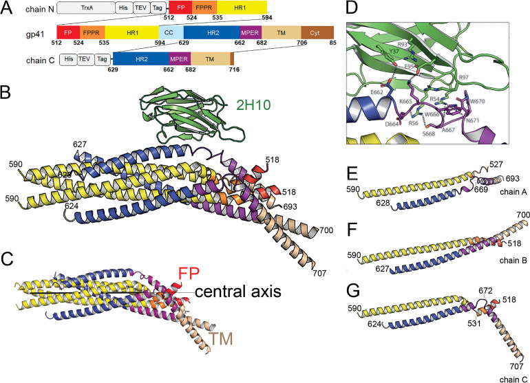
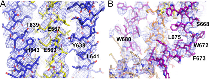
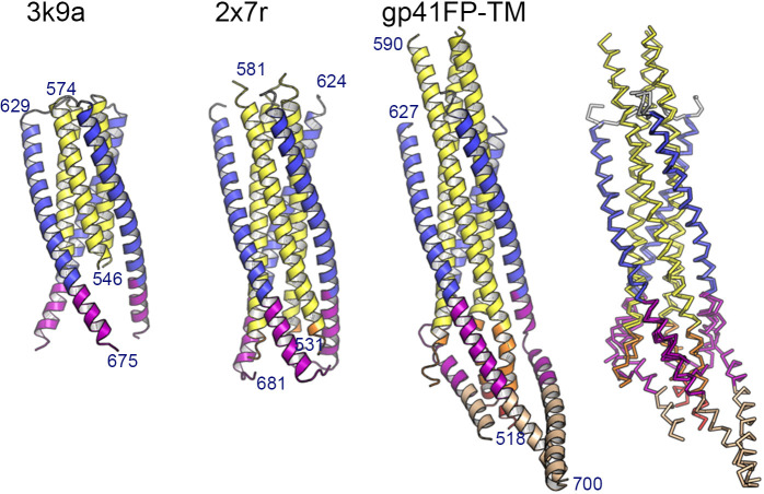
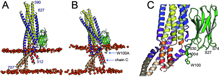
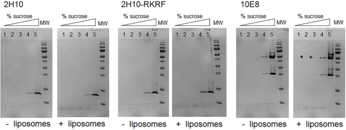
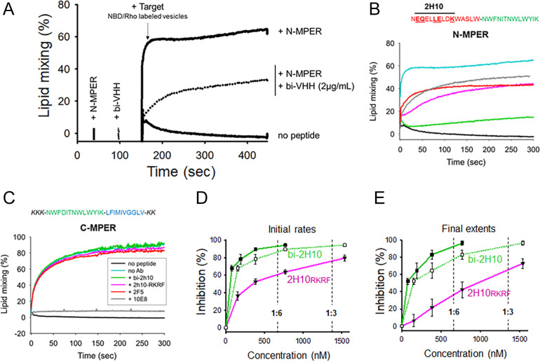
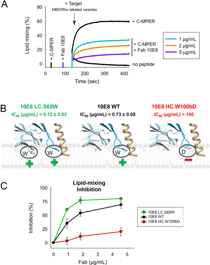
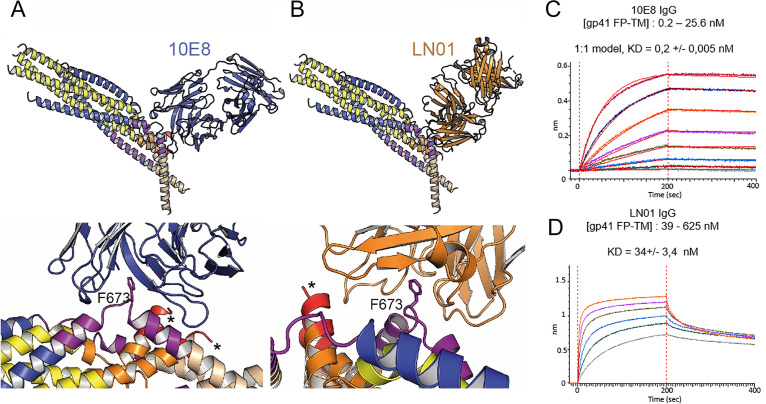
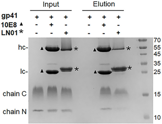
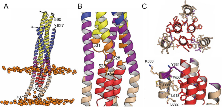
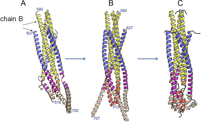
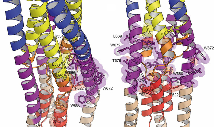
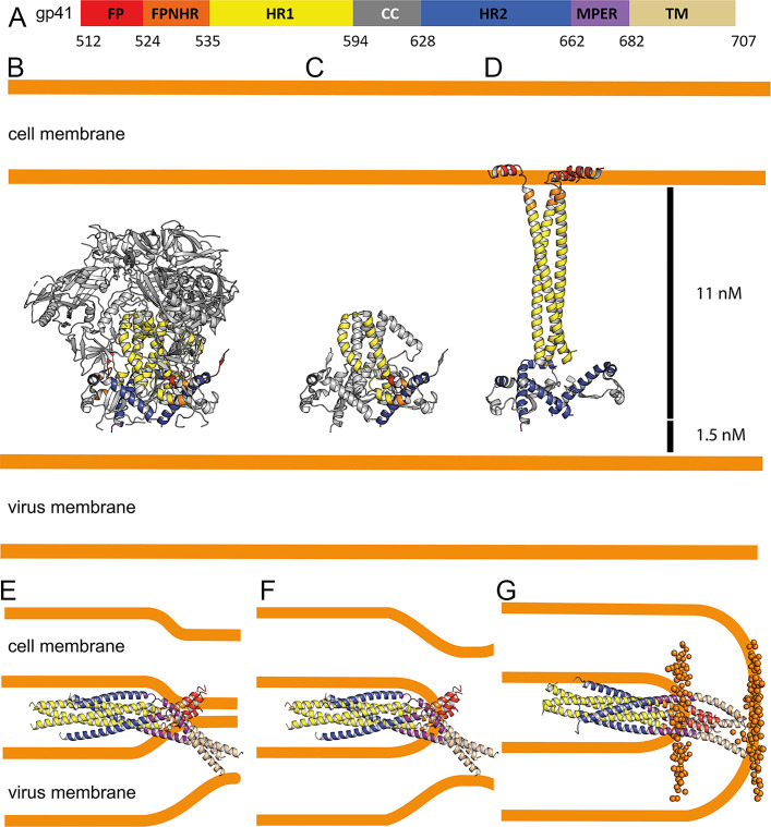
The HIV-1 gp120/gp41 trimer undergoes a series of conformational changes in order to catalyze gp41-induced fusion of viral and cellular membranes. Here, we present the crystal structure of gp41 locked in a fusion intermediate state by an MPER-specific neutralizing antibody. The structure illustrates the conformational plasticity of the six membrane anchors arranged asymmetrically with the fusion peptides and the transmembrane regions pointing into different directions. Hinge regions located adjacent to the fusion peptide and the transmembrane region facilitate the conformational flexibility that allows high-affinity binding of broadly neutralizing anti-MPER antibodies. Molecular dynamics simulation of the MPER Ab-stabilized gp41 conformation reveals a possible transition pathway into the final post-fusion conformation with the central fusion peptides forming a hydrophobic core with flanking transmembrane regions. This suggests that MPER-specific broadly neutralizing antibodies can block final steps of refolding of the fusion peptide and the transmembrane region, which is required for completing membrane fusion.

摘要

HIV-1 gp120/gp41 三聚体发生一系列构象变化,以催化 gp41 诱导的病毒和细胞膜融合。在这里,我们展示了 gp41 被一种 MPER 特异性中和抗体锁定在融合中间状态的晶体结构。该结构说明了六个膜锚定的构象可塑性,它们不对称地排列融合肽和跨膜区域指向不同的方向。位于融合肽和跨膜区域附近的铰链区域促进了构象灵活性,从而允许高亲和力结合广谱中和抗 MPER 抗体。对 MPER Ab 稳定的 gp41 构象的分子动力学模拟揭示了一种可能的过渡途径,进入最终的融合后构象,其中中央融合肽形成一个疏水区,侧翼的跨膜区域。这表明,MPER 特异性广谱中和抗体可以阻止融合肽和跨膜区域的最终折叠,这是完成膜融合所必需的。

https://cdn.ncbi.nlm.nih.gov/pmc/blobs/dfe5/8084527/36ccd522d846/elife-65005-resp-fig3.jpg
https://cdn.ncbi.nlm.nih.gov/pmc/blobs/dfe5/8084527/b87eb51e28a2/elife-65005-fig1.jpg
https://cdn.ncbi.nlm.nih.gov/pmc/blobs/dfe5/8084527/81aca60c0fd8/elife-65005-fig1-figsupp1.jpg
https://cdn.ncbi.nlm.nih.gov/pmc/blobs/dfe5/8084527/a7572d232256/elife-65005-fig1-figsupp2.jpg
https://cdn.ncbi.nlm.nih.gov/pmc/blobs/dfe5/8084527/4f1725177749/elife-65005-fig1-figsupp3.jpg
https://cdn.ncbi.nlm.nih.gov/pmc/blobs/dfe5/8084527/2e9cfd9a6f41/elife-65005-fig1-figsupp4.jpg
https://cdn.ncbi.nlm.nih.gov/pmc/blobs/dfe5/8084527/d623f3c6a623/elife-65005-fig1-figsupp5.jpg
https://cdn.ncbi.nlm.nih.gov/pmc/blobs/dfe5/8084527/331859275012/elife-65005-fig1-figsupp6.jpg
https://cdn.ncbi.nlm.nih.gov/pmc/blobs/dfe5/8084527/e87c7c138de1/elife-65005-fig1-figsupp7.jpg
https://cdn.ncbi.nlm.nih.gov/pmc/blobs/dfe5/8084527/602c74a889f4/elife-65005-fig2.jpg
https://cdn.ncbi.nlm.nih.gov/pmc/blobs/dfe5/8084527/42ab1356e843/elife-65005-fig2-figsupp1.jpg
https://cdn.ncbi.nlm.nih.gov/pmc/blobs/dfe5/8084527/554d97f1ceb5/elife-65005-fig3.jpg
https://cdn.ncbi.nlm.nih.gov/pmc/blobs/dfe5/8084527/71922ba40c14/elife-65005-fig3-figsupp1.jpg
https://cdn.ncbi.nlm.nih.gov/pmc/blobs/dfe5/8084527/cb613f14b636/elife-65005-fig4.jpg
https://cdn.ncbi.nlm.nih.gov/pmc/blobs/dfe5/8084527/e3bdf00d6f24/elife-65005-fig4-figsupp1.jpg
https://cdn.ncbi.nlm.nih.gov/pmc/blobs/dfe5/8084527/152c99ca15b0/elife-65005-fig4-figsupp2.jpg
https://cdn.ncbi.nlm.nih.gov/pmc/blobs/dfe5/8084527/68b1e94154e3/elife-65005-fig5.jpg
https://cdn.ncbi.nlm.nih.gov/pmc/blobs/dfe5/8084527/74f5f8c742cf/elife-65005-resp-fig1.jpg
https://cdn.ncbi.nlm.nih.gov/pmc/blobs/dfe5/8084527/93296938f8df/elife-65005-resp-fig2.jpg
https://cdn.ncbi.nlm.nih.gov/pmc/blobs/dfe5/8084527/36ccd522d846/elife-65005-resp-fig3.jpg

相似文献

1
Structure of HIV-1 gp41 with its membrane anchors targeted by neutralizing antibodies.HIV-1 gp41 结构及其中和抗体靶向的膜锚定。
Elife. 2021 Apr 19;10:e65005. doi: 10.7554/eLife.65005.
2
Cholesterol-dependent membrane fusion induced by the gp41 membrane-proximal external region-transmembrane domain connection suggests a mechanism for broad HIV-1 neutralization.由gp41膜近端外部区域-跨膜结构域连接诱导的胆固醇依赖性膜融合提示了一种广泛中和HIV-1的机制。
J Virol. 2014 Nov;88(22):13367-77. doi: 10.1128/JVI.02151-14. Epub 2014 Sep 10.
3
Effect of diversity in gp41 membrane proximal external region of primary HIV-1 Indian subtype C sequences on interaction with broadly neutralizing antibodies 4E10 and 10E8.原发性 HIV-1 印度 C 亚型 gp41 膜近端外部区多样性对与广泛中和抗体 4E10 和 10E8 相互作用的影响。
Virus Res. 2019 Nov;273:197763. doi: 10.1016/j.virusres.2019.197763. Epub 2019 Sep 22.
4
Crystal structure of HIV-1 gp41 including both fusion peptide and membrane proximal external regions.HIV-1 gp41 包含融合肽和膜近端外部区域的晶体结构。
PLoS Pathog. 2010 May 6;6(5):e1000880. doi: 10.1371/journal.ppat.1000880.
5
Structural Basis for Broad HIV-1 Neutralization by the MPER-Specific Human Broadly Neutralizing Antibody LN01.MPER 特异性人源广谱中和抗体 LN01 广谱中和 HIV-1 的结构基础。
Cell Host Microbe. 2019 Nov 13;26(5):623-637.e8. doi: 10.1016/j.chom.2019.09.016. Epub 2019 Oct 22.
6
Structure-guided alterations of the gp41-directed HIV-1 broadly neutralizing antibody 2F5 reveal new properties regarding its neutralizing function.结构导向的 HIV-1 广谱中和抗体 2F5 的改变揭示了其中和功能的新特性。
PLoS Pathog. 2012;8(7):e1002806. doi: 10.1371/journal.ppat.1002806. Epub 2012 Jul 19.
7
Broadly neutralizing anti-HIV-1 antibodies disrupt a hinge-related function of gp41 at the membrane interface.广泛中和抗HIV-1抗体在膜界面破坏gp41的铰链相关功能。
Proc Natl Acad Sci U S A. 2009 Jun 2;106(22):9057-62. doi: 10.1073/pnas.0901474106. Epub 2009 May 19.
8
HIV type 1 Env precursor cleavage state affects recognition by both neutralizing and nonneutralizing gp41 antibodies.1型人类免疫缺陷病毒包膜前体的切割状态影响中和性和非中和性gp41抗体的识别。
AIDS Res Hum Retroviruses. 2011 Aug;27(8):877-87. doi: 10.1089/AID.2010.0281. Epub 2011 Jan 19.
9
Neutralizing Antibodies Targeting HIV-1 gp41.靶向 HIV-1 gp41 的中和抗体。
Viruses. 2020 Oct 23;12(11):1210. doi: 10.3390/v12111210.
10
Mechanism of HIV-1 neutralization by antibodies targeting a membrane-proximal region of gp41.针对 gp41 膜近端区域的抗体对 HIV-1 的中和作用机制。
J Virol. 2014 Jan;88(2):1249-58. doi: 10.1128/JVI.02664-13. Epub 2013 Nov 13.

引用本文的文献

1
Unveiling the structural spectrum of SARS-CoV-2 fusion by in situ cryo-ET.通过原位冷冻电子断层扫描揭示严重急性呼吸综合征冠状病毒2(SARS-CoV-2)融合的结构谱。
Nat Commun. 2025 Jun 3;16(1):5150. doi: 10.1038/s41467-025-60406-z.
2
Conformational trajectory of the HIV-1 fusion peptide during CD4-induced envelope opening.HIV-1融合肽在CD4诱导包膜开放过程中的构象轨迹。
Nat Commun. 2025 May 17;16(1):4595. doi: 10.1038/s41467-025-59721-2.
3
Current methods for detecting and assessing HIV-1 antibody resistance.检测和评估HIV-1抗体耐药性的当前方法。

本文引用的文献

1
Electron tomography visualization of HIV-1 fusion with target cells using fusion inhibitors to trap the pre-hairpin intermediate.利用融合抑制剂捕获发夹前中间体来可视化 HIV-1 与靶细胞融合的电子断层扫描。
Elife. 2020 Jul 22;9:e58411. doi: 10.7554/eLife.58411.
2
Subnanometer structures of HIV-1 envelope trimers on aldrithiol-2-inactivated virus particles.HIV-1 包膜三聚体在硫代乙醇醛-2 灭活病毒颗粒上的亚纳米结构。
Nat Struct Mol Biol. 2020 Aug;27(8):726-734. doi: 10.1038/s41594-020-0452-2. Epub 2020 Jun 29.
3
Structural transitions in influenza haemagglutinin at membrane fusion pH.
Front Immunol. 2025 Jan 6;15:1443377. doi: 10.3389/fimmu.2024.1443377. eCollection 2024.
4
The Role of Peptides in Combatting HIV Infection: Applications and Insights.肽在抗击 HIV 感染中的作用:应用与见解。
Molecules. 2024 Oct 19;29(20):4951. doi: 10.3390/molecules29204951.
5
A nanobody interaction with SARS-COV-2 Spike allows the versatile targeting of lentivirus vectors.一种与 SARS-COV-2 刺突蛋白相互作用的纳米抗体允许灵活靶向慢病毒载体。
J Virol. 2024 Sep 17;98(9):e0079524. doi: 10.1128/jvi.00795-24. Epub 2024 Aug 29.
6
Insertion and Anchoring of HIV-1 Fusion Peptide into Complex Membrane Mimicking Human T-cell.HIV-1融合肽在模拟人T细胞的复合膜中的插入与锚定
bioRxiv. 2024 Aug 4:2024.08.02.606381. doi: 10.1101/2024.08.02.606381.
7
Molecular Dynamics Investigation of Lipid-Specific Interactions with a Fusion Peptide.分子动力学研究融合肽与脂类的特异性相互作用。
Biomolecules. 2024 Feb 27;14(3):285. doi: 10.3390/biom14030285.
8
Inadequate structural constraint on Fab approach rather than paratope elicitation limits HIV-1 MPER vaccine utility.Fab 进入的结构约束不足而非中和表位诱导限制了 HIV-1 MPER 疫苗的应用。
Nat Commun. 2023 Nov 8;14(1):7218. doi: 10.1038/s41467-023-42097-6.
9
Molecular recognition of a membrane-anchored HIV-1 pan-neutralizing epitope.膜锚定 HIV-1 泛中和表位的分子识别。
Commun Biol. 2022 Nov 18;5(1):1265. doi: 10.1038/s42003-022-04219-6.
10
Membrane attachment and fusion of HIV-1, influenza A, and SARS-CoV-2: resolving the mechanisms with biophysical methods.人类免疫缺陷病毒1型、甲型流感病毒和严重急性呼吸综合征冠状病毒2的膜附着与融合:用生物物理方法解析其机制
Biophys Rev. 2022 Oct 11;14(5):1109-1140. doi: 10.1007/s12551-022-00999-7. eCollection 2022 Oct.
流感血凝素在膜融合 pH 值时的结构转变。
Nature. 2020 Jul;583(7814):150-153. doi: 10.1038/s41586-020-2333-6. Epub 2020 May 27.
4
The Conformational States of the HIV-1 Envelope Glycoproteins.HIV-1 包膜糖蛋白的构象状态。
Trends Microbiol. 2020 Aug;28(8):655-667. doi: 10.1016/j.tim.2020.03.007. Epub 2020 May 14.
5
HIV-1 Envelope and MPER Antibody Structures in Lipid Assemblies.HIV-1 包膜和 MPER 抗体在脂质组装体中的结构。
Cell Rep. 2020 Apr 28;31(4):107583. doi: 10.1016/j.celrep.2020.107583.
6
Structural Basis for Broad HIV-1 Neutralization by the MPER-Specific Human Broadly Neutralizing Antibody LN01.MPER 特异性人源广谱中和抗体 LN01 广谱中和 HIV-1 的结构基础。
Cell Host Microbe. 2019 Nov 13;26(5):623-637.e8. doi: 10.1016/j.chom.2019.09.016. Epub 2019 Oct 22.
7
Molecular Mechanism of HIV-1 Entry.HIV-1 进入的分子机制。
Trends Microbiol. 2019 Oct;27(10):878-891. doi: 10.1016/j.tim.2019.06.002. Epub 2019 Jun 28.
8
Conformational Plasticity in the HIV-1 Fusion Peptide Facilitates Recognition by Broadly Neutralizing Antibodies.HIV-1 融合肽的构象可塑性促进了广谱中和抗体的识别。
Cell Host Microbe. 2019 Jun 12;25(6):873-883.e5. doi: 10.1016/j.chom.2019.04.011.
9
Capturing the inherent structural dynamics of the HIV-1 envelope glycoprotein fusion peptide.捕获 HIV-1 包膜糖蛋白融合肽的固有结构动力学。
Nat Commun. 2019 Feb 15;10(1):763. doi: 10.1038/s41467-019-08738-5.
10
Molecular recognition of the native HIV-1 MPER revealed by STED microscopy of single virions.利用单病毒颗粒的 STED 显微镜观察揭示了 HIV-1 MPER 的天然分子识别。
Nat Commun. 2019 Jan 8;10(1):78. doi: 10.1038/s41467-018-07962-9.