Pang Guodong, Shao Chunchun, Lv Yao, Zhao Fang
Department of Radiology.
Department of Evidence-Based Medicine, The Second Hospital, Cheeloo College of Medicine, Shandong University, Jinan.
Medicine (Baltimore). 2021 Apr 23;100(16):e25627. doi: 10.1097/MD.0000000000025627.
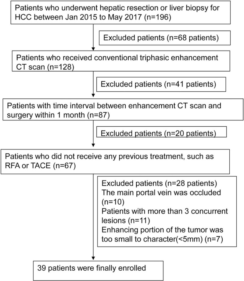
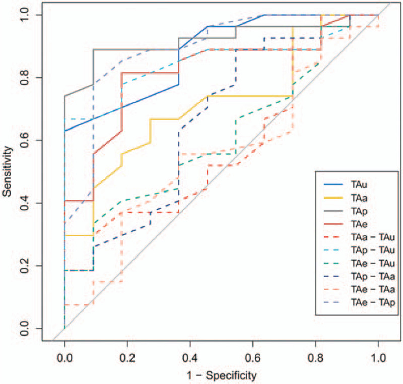
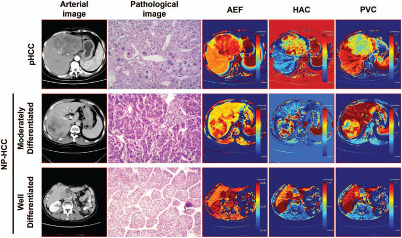
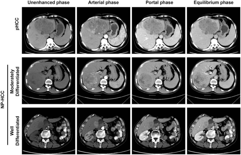
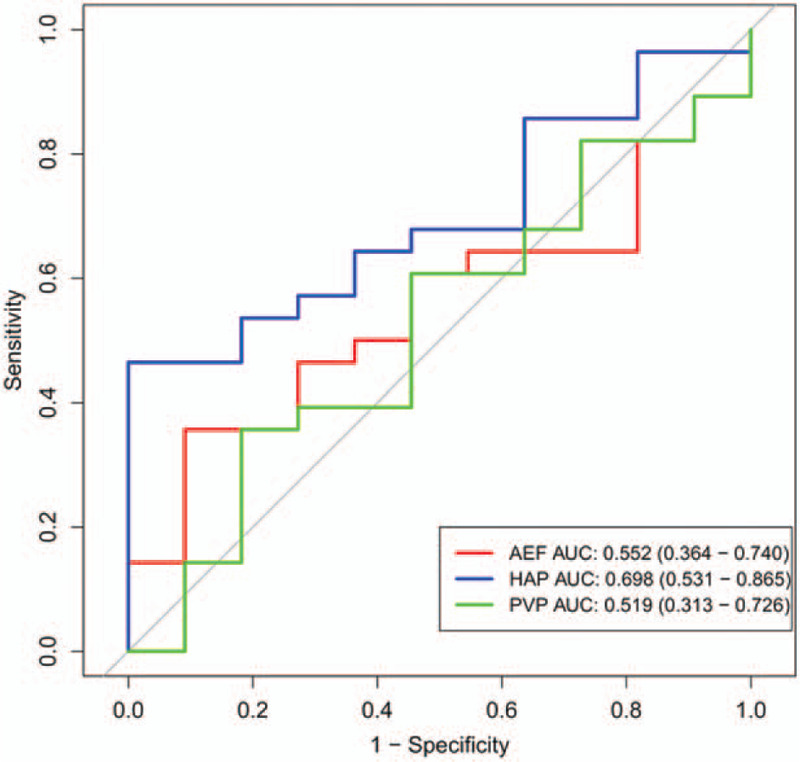
The aim of the current study was to explore the value of tumor attenuation and quantitative analysis of perfusion parameters obtained from traditional tri-phasic CT scans in grading hepatocellular carcinoma (HCC).Totally 39 patients (42 lesion samples) with pathologically confirmed HCC who underwent tri-phasic CT scans were enrolled. HCC lesions were divided into non-poorly differentiated HCC (NP-HCC; n = 31) and poorly differentiated HCC (pHCC; n = 11). All lesions were divided into 5 groups according to the attenuation on different CT enhancement phase. The values of tumor attenuation on different scanning phases were measured. The following parameters were calculated: arterial enhancement fraction (AEF), portal venous supply coefficient (PVC), and hepatic arterial supply coefficient (HAC). The relationship of perfusion parameters with the histological grade of HCC was analyzed. Receiver operating characteristic curves were generated.No significant correlation was observed between the perfusion parameters and tumor grading. Only HAC showed a non-significant trend in different grades of HCC (pHCC < NP-HCC; P = .07). The pHCC cases had significantly decreased values of tumor attenuation on the unenhanced phase (TAu), tumor attenuation on the portal phase portal phase (TAp), and equilibrium phase (TAe) (P < .01). The difference of tumor attenuation between the portal phase and the unenhanced phase (TAp-TAu) of the pHCC cases was decreased than that of the NP-HCC cases (P < .01), whereas the difference of attenuation between the equilibrium phase and portal phase (TAe-TAp) was significantly higher in the pHCC cases than that in the NP-HCC cases (P < .01). TAe-TAp had the highest area under the curve. The number of tumor enhancement pattern in Group 5 of HCCs with a diameter of 3 cm or more was significantly more than that of HCCs with a diameter of less than 3 cm or with other different enhancement patterns (P < .01).Histological HCC grading cannot be predicted by the perfusion parameters derived from traditional tri-phasic CT scans, whereas the tumor attenuation on different phases and the tumor attenuation differences among different phases, especially the mean value of TAe-TAp, might be useful for non-invasive prediction on the degree of HCC differentiation.
本研究的目的是探讨传统三期CT扫描获得的肿瘤衰减值及灌注参数定量分析在肝细胞癌(HCC)分级中的价值。共纳入39例经病理证实为HCC且接受过三期CT扫描的患者(42个病灶样本)。HCC病灶分为非低分化HCC(NP-HCC;n = 31)和低分化HCC(pHCC;n = 11)。所有病灶根据不同CT增强期的衰减情况分为5组。测量不同扫描期的肿瘤衰减值。计算以下参数:动脉增强分数(AEF)、门静脉供血系数(PVC)和肝动脉供血系数(HAC)。分析灌注参数与HCC组织学分级的关系。绘制受试者工作特征曲线。未观察到灌注参数与肿瘤分级之间存在显著相关性。仅HAC在不同分级的HCC中显示出不显著的趋势(pHCC < NP-HCC;P = 0.07)。pHCC病例在平扫期(TAu)、门静脉期(TAp)和平衡期(TAe)的肿瘤衰减值显著降低(P < 0.01)。pHCC病例门静脉期与平扫期的肿瘤衰减差值(TAp-TAu)低于NP-HCC病例(P < 0.01),而pHCC病例平衡期与门静脉期的衰减差值(TAe-TAp)显著高于NP-HCC病例(P < 0.01)。TAe-TAp的曲线下面积最大。直径≥3 cm的HCC第5组肿瘤强化模式的数量显著多于直径<3 cm或具有其他不同强化模式的HCC(P < 0.01)。传统三期CT扫描得出的灌注参数无法预测HCC的组织学分级,而不同期的肿瘤衰减值及不同期之间的肿瘤衰减差值,尤其是TAe-TAp的平均值,可能有助于对HCC分化程度进行无创预测。