R&D Center, OncoInsight Co. Ltd, Seoul, South Korea.
Laboratory of Stem Cell, NEXEL, Seoul, South Korea.
Arthritis Res Ther. 2021 Apr 21;23(1):124. doi: 10.1186/s13075-021-02491-1.
In the pathogenesis of rheumatoid arthritis (RA), the role of mast cells has not been revealed clearly. We aimed to define the inflammatory and tissue-destructive roles of mast cells in rheumatoid arthritis (RA).
Serum and synovial fluid (SF) concentration levels of tryptase, chymase, and histamine were quantified using ELISA. After activating mast cells using IL-33, the production of TNF-α, IL-1β, IL-6, IL-17, RANKL, and MMPs was determined using real-time PCR and ELISA. Osteoclastogenesis was assessed in CD14+ monocytes from peripheral blood and SF, which were cultured with IL-33-activated mast cells, by counting TRAP-positive multinucleated cells.
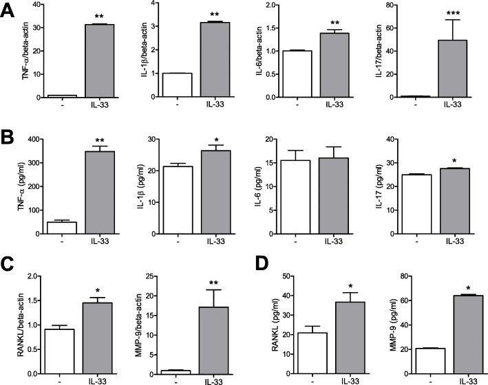
The concentration levels of serum tryptase, chymase, and histamine and SF histamine were higher in patients with RA than in controls. FcεR1 and c-kit-positive mast cells were higher in RA synovium than in osteoarthritic (OA) synovium. Stimulation of mast cells by IL-33 increased the number of trypatse+chymase- and tryptase+chymase+ mast cells. IL-33 stimulation also increased the gene expression levels of TNF-α, IL-1β, IL-6, IL-17, RANKL, and MMP-9 in mast cells. Furthermore, IL-33 stimulated human CD14+ monocytes to differentiate into TRAP+ multinucleated osteoclasts. When CD14+ monocytes were co-cultured with mast cells, osteoclast differentiation was increased. Additionally, IL-33-activated mast cells stimulated osteoclast differentiation. The inhibition of intercellular contact between mast cells and monocytes using inserts reduced osteoclast differentiation.
IL-33 increased inflammatory and tissue-destructive cytokines by activation of mast cells. Mast cells stimulated osteoclast differentiation in monocytes. Mast cells could stimulate osteoclastogenesis indirectly through production of tissue-destructive cytokines and directly through stimulation of osteoclast precursors.
在类风湿关节炎(RA)的发病机制中,肥大细胞的作用尚未明确。本研究旨在明确肥大细胞在类风湿关节炎(RA)中的炎症和组织破坏作用。
采用 ELISA 法检测血清和滑膜液(SF)中类胰蛋白酶、糜蛋白酶和组胺的浓度。用白细胞介素-33(IL-33)激活肥大细胞后,采用实时 PCR 和 ELISA 法检测 TNF-α、IL-1β、IL-6、IL-17、RANKL 和 MMP 的产生。用 IL-33 激活的肥大细胞与外周血和 SF 中的 CD14+单核细胞共培养,通过计数 TRAP 阳性多核细胞来评估破骨细胞生成。
与对照组相比,RA 患者的血清类胰蛋白酶、糜蛋白酶和组胺浓度以及 SF 中的组胺浓度更高。RA 滑膜中的 FcεR1 和 c-kit 阳性肥大细胞多于骨关节炎(OA)滑膜。IL-33 刺激肥大细胞增加了 tryptase+chymase-和 tryptase+chymase+肥大细胞的数量。IL-33 刺激还增加了肥大细胞中 TNF-α、IL-1β、IL-6、IL-17、RANKL 和 MMP-9 的基因表达水平。此外,IL-33 刺激人 CD14+单核细胞分化为 TRAP+多核破骨细胞。当 CD14+单核细胞与肥大细胞共培养时,破骨细胞分化增加。此外,IL-33 激活的肥大细胞刺激破骨细胞分化。使用插入物抑制肥大细胞与单核细胞之间的细胞间接触减少了破骨细胞分化。
IL-33 通过激活肥大细胞增加了炎症和组织破坏性细胞因子。肥大细胞刺激单核细胞中的破骨细胞分化。肥大细胞可以通过产生组织破坏性细胞因子和直接刺激破骨细胞前体间接刺激破骨细胞生成。