Africa Health Research Institute (AHRI), Durban, South Africa.
School of Laboratory Medicine and Medical Sciences, University of KwaZulu-Natal, Durban, South Africa.
Front Immunol. 2021 Apr 9;12:631410. doi: 10.3389/fimmu.2021.631410. eCollection 2021.
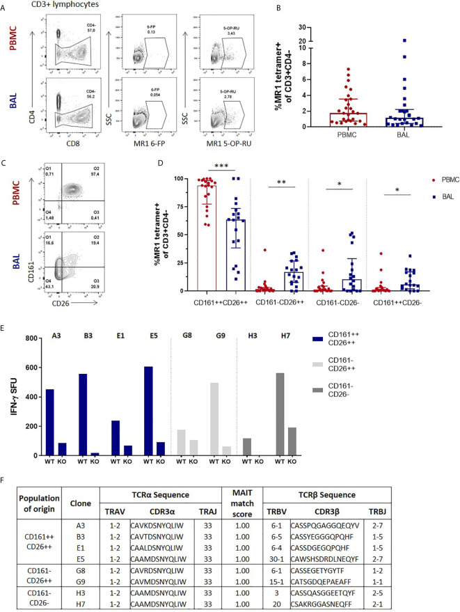
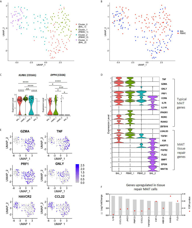
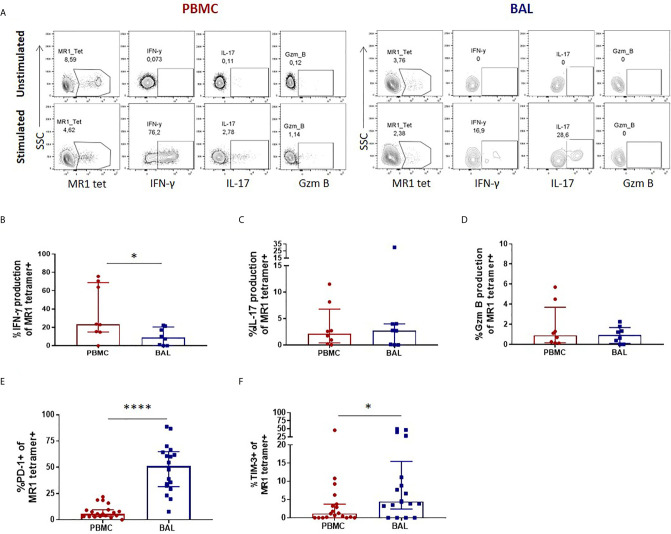
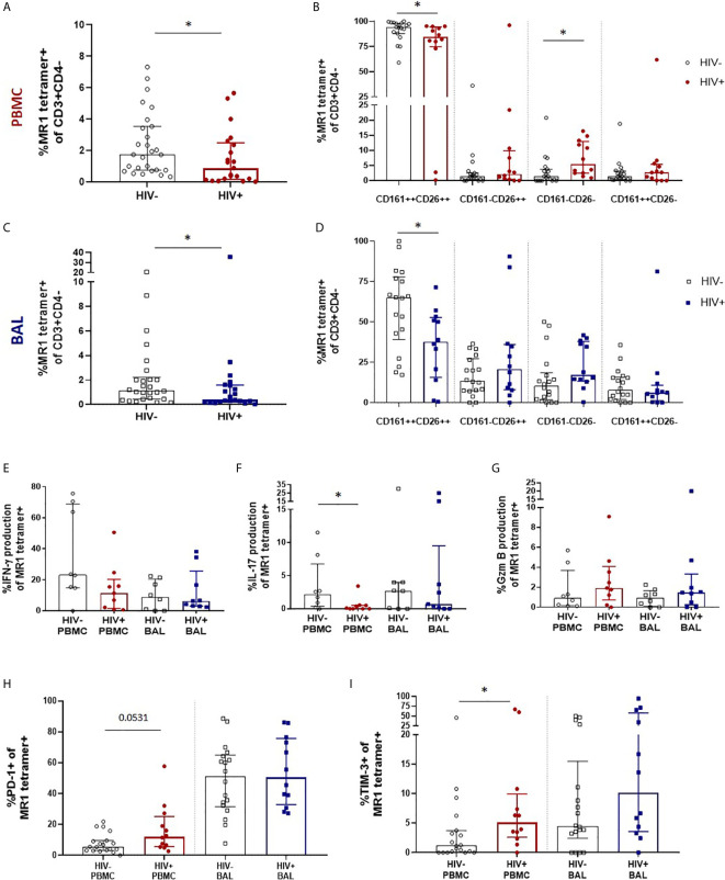
Mucosal associated invariant T (MAIT) cells are a class of innate-like T cells that utilize a semi-invariant αβ T cell receptor to recognize small molecule ligands produced by bacteria and fungi. Despite growing evidence that immune cells at mucosal surfaces are often phenotypically and functionally distinct from those in the peripheral circulation, knowledge about the characteristics of MAIT cells at the lung mucosal surface, the site of exposure to respiratory pathogens, is limited. HIV infection has been shown to have a profound effect on the number and function of MAIT cells in the peripheral blood, but its effect on lung mucosal MAIT cells is unknown. We examined the phenotypic, functional, and transcriptomic features of major histocompatibility complex (MHC) class I-related (MR1)-restricted MAIT cells from the peripheral blood and bronchoalveolar compartments of otherwise healthy individuals with latent () infection who were either HIV uninfected or HIV infected. Peripheral blood MAIT cells consistently co-expressed typical MAIT cell surface markers CD161 and CD26 in HIV-negative individuals, while paired bronchoalveolar MAIT cells displayed heterogenous expression of these markers. Bronchoalveolar MAIT cells produced lower levels of pro-inflammatory cytokine IFN-γ and expressed higher levels of co-inhibitory markers PD-1 and TIM-3 than peripheral MAIT cells. HIV infection resulted in decreased frequencies and pro-inflammatory function of peripheral blood MAIT cells, while in the bronchoalveolar compartment MAIT cell frequency was decreased but phenotype and function were not significantly altered. Single-cell transcriptomic analysis demonstrated greater heterogeneity among bronchoalveolar compared to peripheral blood MAIT cells and suggested a distinct subset in the bronchoalveolar compartment. The transcriptional features of this bronchoalveolar subset were associated with MAIT cell tissue repair functions. In summary, we found previously undescribed phenotypic and transcriptional heterogeneity of bronchoalveolar MAIT cells in HIV-negative people. In HIV infection, we found numeric depletion of MAIT cells in both anatomical compartments but preservation of the novel phenotypic and transcriptional features of bronchoalveolar MAIT cells.
黏膜相关不变 T(MAIT)细胞是一类先天样 T 细胞,它们利用半不变的αβ T 细胞受体来识别细菌和真菌产生的小分子配体。尽管越来越多的证据表明,黏膜表面的免疫细胞在表型和功能上通常与外周循环中的免疫细胞不同,但对于肺部黏膜表面(即暴露于呼吸道病原体的部位)MAIT 细胞的特征知之甚少。HIV 感染已被证明对外周血中 MAIT 细胞的数量和功能有深远影响,但对肺部黏膜 MAIT 细胞的影响尚不清楚。我们研究了潜伏性()感染的、无 HIV 感染或 HIV 感染的健康个体外周血和支气管肺泡腔中主要组织相容性复合体(MHC)I 类相关(MR1)限制的 MAIT 细胞的表型、功能和转录组特征。在 HIV 阴性个体中,外周血 MAIT 细胞一致表达典型的 MAIT 细胞表面标记物 CD161 和 CD26,而配对的支气管肺泡 MAIT 细胞则表现出这些标记物的异质性表达。支气管肺泡 MAIT 细胞产生的促炎细胞因子 IFN-γ 水平较低,共抑制标记物 PD-1 和 TIM-3 的表达水平较高。HIV 感染导致外周血 MAIT 细胞频率降低和促炎功能受损,而在支气管肺泡腔中,MAIT 细胞频率降低,但表型和功能没有明显改变。单细胞转录组分析表明,与外周血 MAIT 细胞相比,支气管肺泡 MAIT 细胞的异质性更大,并提示支气管肺泡腔中有一个独特的亚群。该支气管肺泡亚群的转录特征与 MAIT 细胞的组织修复功能相关。总之,我们发现了 HIV 阴性人群中以前未描述的支气管肺泡 MAIT 细胞的表型和转录异质性。在 HIV 感染中,我们发现两个解剖部位的 MAIT 细胞数量均减少,但保留了支气管肺泡 MAIT 细胞的新型表型和转录特征。