McCook Oscar, Scheuerle Angelika, Denoix Nicole, Kapapa Thomas, Radermacher Peter, Merz Tamara
Institute for Anesthesiological Pathophysiology and Process Engineering, Ulm University Medical Center, Ulm, Germany.
Department of Neuropathology, Ulm University Medical Center, Günzburg, Germany.
Neural Regen Res. 2021 Dec;16(12):2376-2382. doi: 10.4103/1673-5374.313018.
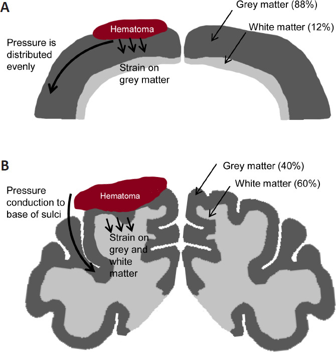
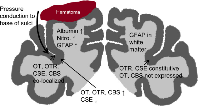
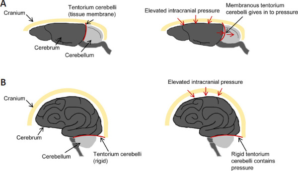
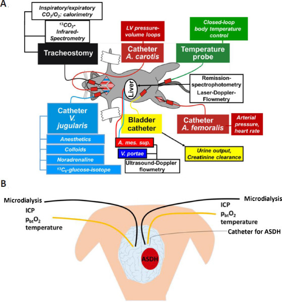
In the porcine model discussed in this review, the acute subdural hematoma was induced by subdural injection of autologous blood over the left parietal cortex, which led to a transient elevation of the intracerebral pressure, measured by bilateral neuromonitoring. The hematoma-induced brain injury was associated with albumin extravasation, oxidative stress, reactive astrogliosis and microglial activation in the ipsilateral hemisphere. Further proteins and injury markers were validated to be used for immunohistochemistry of porcine brain tissue. The cerebral expression patterns of oxytocin, oxytocin receptor, cystathionine-γ-lyase and cystathionine-β-synthase were particularly interesting: these four proteins all co-localized at the base of the sulci, where pressure-induced brain injury elicits maximum stress. In this context, the pig is a very relevant translational model in contrast to the rodent brain. The structure of the porcine brain is very similar to the human: the presence of gyri and sulci (gyrencephalic brain), white matter to grey matter proportion and tentorium cerebelli. Thus, pressure-induced injury in the porcine brain, unlike in the rodent brain, is reflective of the human pathophysiology.
在本综述所讨论的猪模型中,通过在左侧顶叶皮质硬膜下注射自体血诱导急性硬膜下血肿,通过双侧神经监测测量发现这导致了颅内压的短暂升高。血肿诱导的脑损伤与同侧半球的白蛋白外渗、氧化应激、反应性星形胶质细胞增生和小胶质细胞激活有关。进一步验证了用于猪脑组织免疫组织化学的蛋白质和损伤标志物。催产素、催产素受体、胱硫醚-γ-裂解酶和胱硫醚-β-合酶的脑表达模式特别有趣:这四种蛋白质都共定位于脑沟底部,压力诱导的脑损伤在那里引发最大应激。在这种情况下,与啮齿动物脑相比,猪是一个非常相关的转化模型。猪脑的结构与人类非常相似:存在脑回和脑沟(脑回脑)、白质与灰质比例以及小脑幕。因此,与啮齿动物脑不同,猪脑中压力诱导的损伤反映了人类病理生理学。