• 文献检索
  • 文档翻译
  • 深度研究
  • 学术资讯
  • Suppr Zotero 插件Zotero 插件
  • 邀请有礼
  • 套餐&价格
  • 历史记录
应用&插件
Suppr Zotero 插件Zotero 插件浏览器插件Mac 客户端Windows 客户端微信小程序
定价
高级版会员购买积分包购买API积分包
服务
文献检索文档翻译深度研究API 文档MCP 服务
关于我们
关于 Suppr公司介绍联系我们用户协议隐私条款
关注我们

Suppr 超能文献

核心技术专利:CN118964589B侵权必究
粤ICP备2023148730 号-1Suppr @ 2026

文献检索

告别复杂PubMed语法,用中文像聊天一样搜索,搜遍4000万医学文献。AI智能推荐,让科研检索更轻松。

立即免费搜索

文件翻译

保留排版,准确专业,支持PDF/Word/PPT等文件格式,支持 12+语言互译。

免费翻译文档

深度研究

AI帮你快速写综述,25分钟生成高质量综述,智能提取关键信息,辅助科研写作。

立即免费体验

应用锥形束 CT 分析颞下颌关节的新方法。

New Method for Analysis of the Temporomandibular Joint Using Cone Beam Computed Tomography.

机构信息

GIG Research Institute, 40-166 Katowice, Poland.

Department of Temporomandibular Disorders, Medical University of Silesia, 41-800 Zabrze, Poland.

出版信息

Sensors (Basel). 2021 Apr 28;21(9):3070. doi: 10.3390/s21093070.

DOI:10.3390/s21093070
PMID:33924981
原文链接:https://pmc.ncbi.nlm.nih.gov/articles/PMC8125202/
Abstract

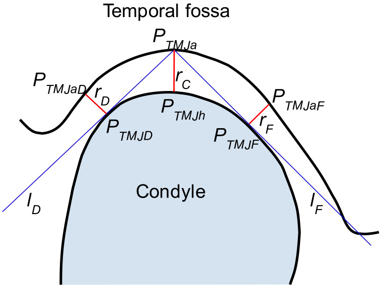
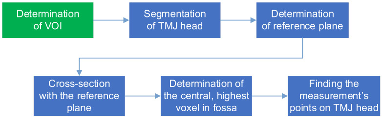
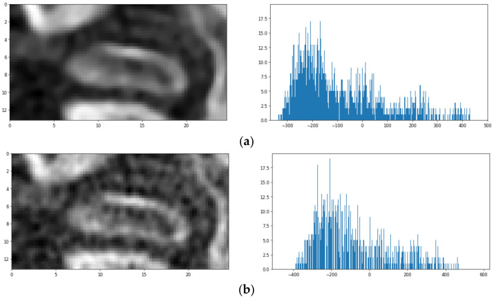
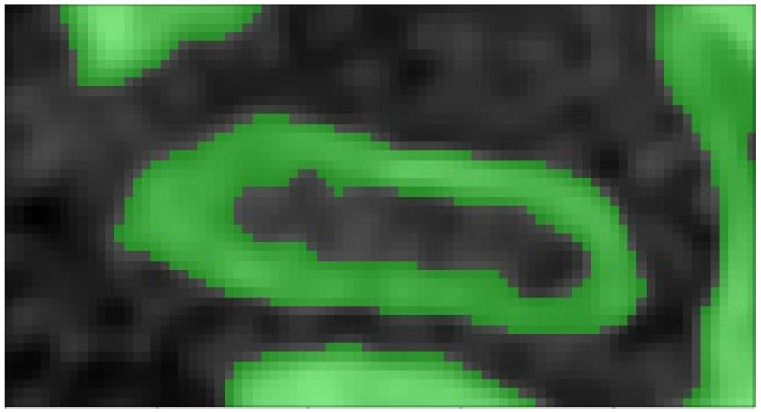
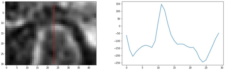
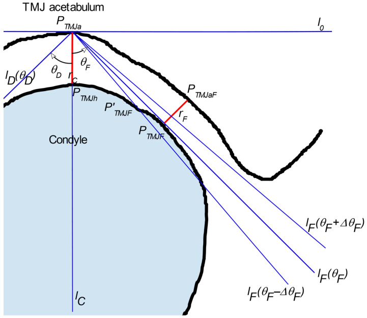
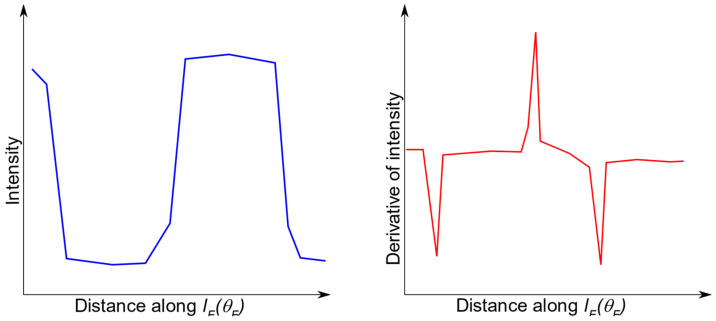
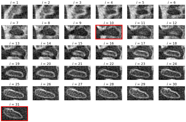
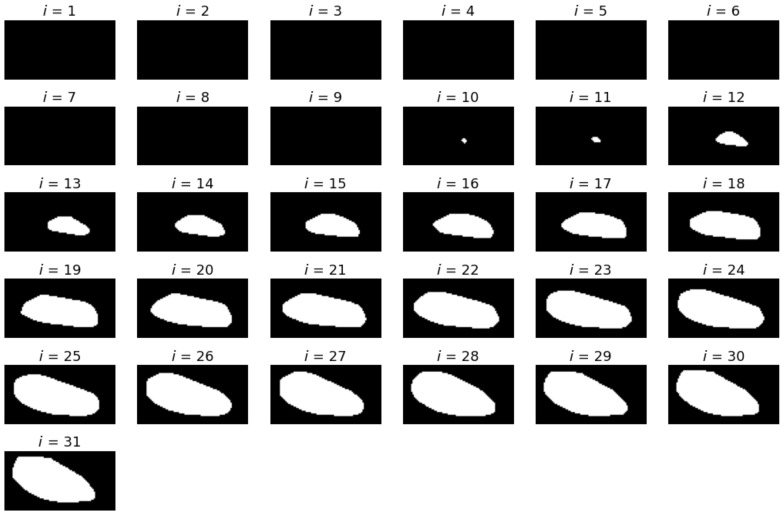
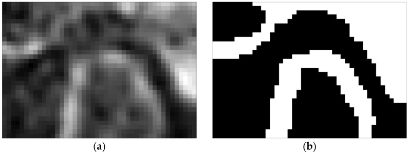
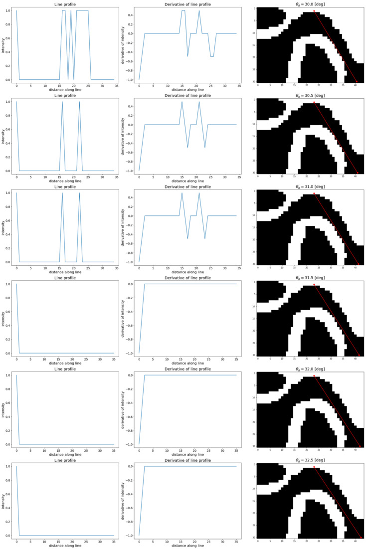
Modern dentistry commonly uses a variety of imaging methods to support diagnosis and treatment. Among them, cone beam computed tomography (CBCT) is particularly useful in presenting head structures, such as the temporomandibular joint (TMJ). The determination of the morphology of the joint is an important part of the diagnosis as well as the monitoring of the treatment results. It can be accomplished by measurement of the TMJ gap width at three selected places, taken at a specific cross-section. This study presents a new approach to these measurements. First, the CBCT images are denoised using curvilinear methods, and the volume of interest is determined. Then, the orientation of the vertical cross-section plane is computed based on segmented axial sections of the TMJ head. Finally, the cross-section plane is used to determine the standardized locations, at which the width of the gap between condyle and fossa is measured. The elaborated method was tested on selected TMJ CBCT scans with satisfactory results. The proposed solution lays the basis for the development of an autonomous method of TMJ index identification.

摘要

现代牙科通常使用多种成像方法来支持诊断和治疗。其中,锥形束计算机断层扫描(CBCT)在呈现头部结构方面特别有用,例如颞下颌关节(TMJ)。关节形态的确定是诊断和监测治疗结果的重要部分。可以通过在特定横截面上取三个选定位置测量 TMJ 间隙宽度来完成。本研究提出了一种新的测量方法。首先,使用曲线方法对 CBCT 图像进行去噪,并确定感兴趣的体积。然后,根据 TMJ 头部的分段轴位图像计算垂直横截平面的方向。最后,使用横截面平面确定测量髁突和窝之间间隙宽度的标准化位置。该详细方法在选定的 TMJ CBCT 扫描上进行了测试,结果令人满意。所提出的解决方案为开发自主的 TMJ 指数识别方法奠定了基础。

https://cdn.ncbi.nlm.nih.gov/pmc/blobs/278a/8125202/7286854aa050/sensors-21-03070-g013.jpg
https://cdn.ncbi.nlm.nih.gov/pmc/blobs/278a/8125202/cb4486a05eee/sensors-21-03070-g001.jpg
https://cdn.ncbi.nlm.nih.gov/pmc/blobs/278a/8125202/733eb2bea829/sensors-21-03070-g002.jpg
https://cdn.ncbi.nlm.nih.gov/pmc/blobs/278a/8125202/c0d59adfdf0a/sensors-21-03070-g003.jpg
https://cdn.ncbi.nlm.nih.gov/pmc/blobs/278a/8125202/554b855c05c9/sensors-21-03070-g004.jpg
https://cdn.ncbi.nlm.nih.gov/pmc/blobs/278a/8125202/97791721c187/sensors-21-03070-g005.jpg
https://cdn.ncbi.nlm.nih.gov/pmc/blobs/278a/8125202/3b666b290d8e/sensors-21-03070-g006.jpg
https://cdn.ncbi.nlm.nih.gov/pmc/blobs/278a/8125202/fb7d52520110/sensors-21-03070-g007.jpg
https://cdn.ncbi.nlm.nih.gov/pmc/blobs/278a/8125202/e954a39a4305/sensors-21-03070-g008.jpg
https://cdn.ncbi.nlm.nih.gov/pmc/blobs/278a/8125202/9336874eeb0a/sensors-21-03070-g009.jpg
https://cdn.ncbi.nlm.nih.gov/pmc/blobs/278a/8125202/9764ec1abd14/sensors-21-03070-g010.jpg
https://cdn.ncbi.nlm.nih.gov/pmc/blobs/278a/8125202/685f54c83172/sensors-21-03070-g011.jpg
https://cdn.ncbi.nlm.nih.gov/pmc/blobs/278a/8125202/6c5f24e831df/sensors-21-03070-g012.jpg
https://cdn.ncbi.nlm.nih.gov/pmc/blobs/278a/8125202/7286854aa050/sensors-21-03070-g013.jpg

相似文献

1
New Method for Analysis of the Temporomandibular Joint Using Cone Beam Computed Tomography.应用锥形束 CT 分析颞下颌关节的新方法。
Sensors (Basel). 2021 Apr 28;21(9):3070. doi: 10.3390/s21093070.
2
The effect of myotonic dystrophy type 1 on temporomandibular joint and dentofacial morphology: A CBCT analysis.1 型肌强直性营养不良对颞下颌关节和牙颌面形态的影响:CBCT 分析。
J Oral Rehabil. 2023 Oct;50(10):958-964. doi: 10.1111/joor.13533. Epub 2023 Jun 16.
3
Comparative cone-beam computed tomography evaluation of the osseous morphology of the temporomandibular joint in temporomandibular dysfunction patients and asymptomatic individuals.颞下颌关节紊乱病患者与无症状个体颞下颌关节骨形态的锥束计算机断层扫描对比评估
Oral Radiol. 2018 Jan;34(1):31-39. doi: 10.1007/s11282-017-0279-7. Epub 2017 Mar 16.
4
Quantification of temporomandibular joint space in patients with juvenile idiopathic arthritis assessed by cone beam computerized tomography.通过锥形束计算机断层扫描评估青少年特发性关节炎患者颞下颌关节间隙的量化分析。
Orthod Craniofac Res. 2024 Apr;27(2):203-210. doi: 10.1111/ocr.12702. Epub 2023 Aug 1.
5
Deep learning for automated segmentation of the temporomandibular joint.用于颞下颌关节自动分割的深度学习
J Dent. 2023 May;132:104475. doi: 10.1016/j.jdent.2023.104475. Epub 2023 Mar 2.
6
Comparative Analysis of Bilateral Temporomandibular Joints in Patients With Unilateral Temporomandibular Joint Complaints Using Cone Beam Computed Tomography.使用锥形束计算机断层扫描对单侧颞下颌关节疾病患者双侧颞下颌关节的比较分析
J Craniofac Surg. 2015 Nov;26(8):e773-6. doi: 10.1097/SCS.0000000000002253.
7
Accuracy of cone-beam computed tomography imaging of the temporomandibular joint: comparisons with panoramic radiology and linear tomography.锥形束计算机断层扫描对颞下颌关节成像的准确性:与全景放射摄影和线性断层扫描的比较。
Am J Orthod Dentofacial Orthop. 2007 Oct;132(4):429-38. doi: 10.1016/j.ajodo.2005.10.032.
8
Bony deviations revealed by cone beam computed tomography of the temporomandibular joint in subjects without ongoing pain.无持续性疼痛患者的颞下颌关节锥形束 CT 检查所显示的骨性偏斜。
J Oral Facial Pain Headache. 2014 Fall;28(4):331-7. doi: 10.11607/ofph.1255.
9
[A preliminary study on the registration of MRI and cone beam CT images of temporomandibular joint disc].颞下颌关节盘MRI与锥形束CT图像配准的初步研究
Zhonghua Kou Qiang Yi Xue Za Zhi. 2020 Oct 9;55(10):772-777. doi: 10.3760/cma.j.cn112144-20200605-00319.
10
Developing a universal and reliable temporomandibular joint reference system for quantifying morphological and positional changes.为了量化形态和位置变化,开发一个通用且可靠的颞下颌关节参考系统。
Eur J Orthod. 2024 Jan 1;46(1). doi: 10.1093/ejo/cjad055.

引用本文的文献

1
Three-dimensional visualization of temporomandibular joint after occlusal changes using facial scanning techniques combined with single cone beam computed tomography.使用面部扫描技术结合单锥束计算机断层扫描对咬合变化后颞下颌关节进行三维可视化。
Clin Oral Investig. 2025 Apr 8;29(5):230. doi: 10.1007/s00784-025-06299-0.
2
A Deep Learning Approach to Intrusion Detection and Segmentation in Pellet Fuels Using Microscopic Images.一种利用微观图像对颗粒燃料中的入侵进行检测和分割的深度学习方法。
Sensors (Basel). 2023 Jul 18;23(14):6488. doi: 10.3390/s23146488.
3
Intra-Articular Injections into the Inferior versus Superior Compartment of the Temporomandibular Joint: A Systematic Review and Meta-Analysis.

本文引用的文献

1
Morphometric analysis of the mandibular fossa in dentate and edentulous patients: A cone beam computed tomography study.有牙和无牙患者下颌窝的形态计量分析:锥形束 CT 研究。
J Prosthet Dent. 2021 May;125(5):758.e1-758.e7. doi: 10.1016/j.prosdent.2021.01.014. Epub 2021 Feb 17.
2
Long-term follow-up of intersegmental displacement after orthognathic surgery using cone-beam computed tomographic superimposition.应用锥形束 CT 叠加技术对正颌手术后节段间位移的长期随访
Angle Orthod. 2020 Jul 1;90(4):548-555. doi: 10.2319/061119-398.
3
Temporomandibular disc displacement with reduction treated with anterior repositioning splint: a 2-year clinical and magnetic resonance imaging (MRI) follow-up.
颞下颌关节下腔与上腔关节内注射:一项系统评价和荟萃分析
J Clin Med. 2023 Feb 19;12(4):1664. doi: 10.3390/jcm12041664.
4
Repeatability of the Vibroarthrogram in the Temporomandibular Joints.颞下颌关节震音图的可重复性。
Sensors (Basel). 2022 Dec 6;22(23):9542. doi: 10.3390/s22239542.
5
Differential Diagnosis of Cysts and Granulomas Supported by Texture Analysis of Intraoral Radiographs.基于口腔内射线照片纹理分析的囊肿和肉芽肿的鉴别诊断。
Sensors (Basel). 2021 Nov 10;21(22):7481. doi: 10.3390/s21227481.
可复性髁突前移位伴关节盘前移位的治疗:2 年的临床和磁共振成像(MRI)随访。
J Biol Regul Homeost Agents. 2020 Jan-Feb;34(1 Suppl. 1):151-160. DENTAL SUPPLEMENT.
4
Symmetry of root and root canal morphology of mandibular incisors: A cone-beam computed tomography study .下颌切牙牙根及根管形态的对称性:一项锥形束计算机断层扫描研究
J Clin Exp Dent. 2019 Jun 1;11(6):e527-e533. doi: 10.4317/jced.55629. eCollection 2019 Jun.
5
A Full-Digital Technique to Mount a Maxillary Arch Scan on a Virtual Articulator.一种在虚拟牙合架上安装上颌弓扫描的全数字化技术。
J Prosthodont. 2019 Mar;28(3):335-338. doi: 10.1111/jopr.13023. Epub 2019 Jan 31.
6
Cone-beam computed tomographic analysis on root and canal morphology of mandibular first permanent molar among multiracial population in Western European population.西欧多民族人群下颌第一恒磨牙牙根及根管形态的锥形束计算机断层扫描分析
Eur J Dent. 2018 Jul-Sep;12(3):434-438. doi: 10.4103/ejd.ejd_116_18.
7
Natural head position: An overview.自然头位:综述。
J Pharm Bioallied Sci. 2015 Aug;7(Suppl 2):S424-7. doi: 10.4103/0975-7406.163488.
8
Technical aspects of dental CBCT: state of the art.牙科锥形束计算机断层扫描的技术层面:当前技术水平
Dentomaxillofac Radiol. 2015;44(1):20140224. doi: 10.1259/dmfr.20140224.
9
The virtual patient in dental medicine.牙科医学中的虚拟患者。
Clin Oral Implants Res. 2015 Jun;26(6):725-6. doi: 10.1111/clr.12379. Epub 2014 Mar 26.
10
Segmentation of Trabecular Jaw Bone on Cone Beam CT Datasets.锥形束CT数据集上的下颌骨小梁分割
Clin Implant Dent Relat Res. 2015 Dec;17(6):1082-91. doi: 10.1111/cid.12217. Epub 2014 Mar 14.