Ag Seleci Didem, Maurer Viktor, Barlas Firat Baris, Porsiel Julian Cedric, Temel Bilal, Ceylan Elcin, Timur Suna, Stahl Frank, Scheper Thomas, Garnweitner Georg
Institute for Particle Technology (iPAT), Technische Universität Braunschweig, 38104 Braunschweig, Germany.
Center of Pharmaceutical Engineering (PVZ), Technische Universität Braunschweig, 38106 Braunschweig, Germany.
Int J Mol Sci. 2021 Apr 27;22(9):4556. doi: 10.3390/ijms22094556.
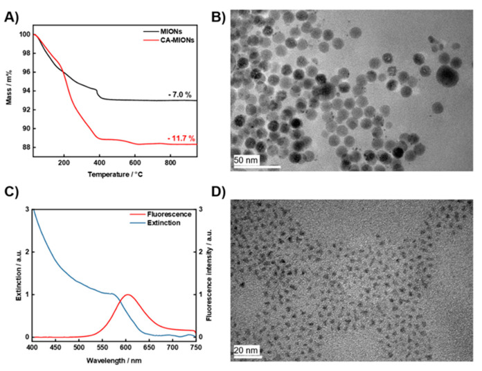
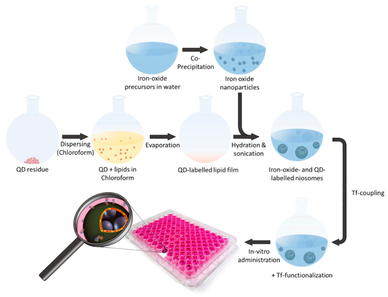
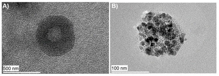
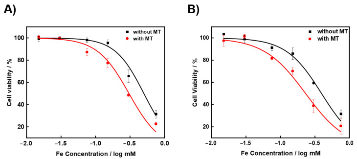
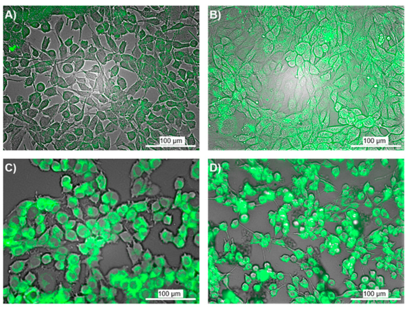
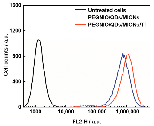
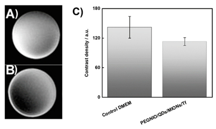
The development of multifunctional nanoscale systems that can mediate efficient tumor targeting, together with high cellular internalization, is crucial for the diagnosis of glioma. The combination of imaging agents into one platform provides dual imaging and allows further surface modification with targeting ligands for specific glioma detection. Herein, transferrin (Tf)-decorated niosomes with integrated magnetic iron oxide nanoparticles (MIONs) and quantum dots (QDs) were formulated (PEGNIO/QDs/MIONs/Tf) for efficient imaging of glioma, supported by magnetic and active targeting. Transmission electron microscopy confirmed the complete co-encapsulation of MIONs and QDs in the niosomes. Flow cytometry analysis demonstrated enhanced cellular uptake of the niosomal formulation by glioma cells. In vitro imaging studies showed that PEGNIO/QDs/MIONs/Tf produces an obvious negative-contrast enhancement effect on glioma cells by magnetic resonance imaging (MRI) and also improved fluorescence intensity under fluorescence microscopy. This novel platform represents the first niosome-based system which combines magnetic nanoparticles and QDs, and has application potential in dual-targeted imaging of glioma.
能够介导高效肿瘤靶向以及高细胞内化的多功能纳米级系统的开发,对于神经胶质瘤的诊断至关重要。将成像剂组合到一个平台中可提供双重成像,并允许用靶向配体进行进一步的表面修饰,以用于特定神经胶质瘤的检测。在此,制备了具有整合的磁性氧化铁纳米颗粒(MIONs)和量子点(QDs)的转铁蛋白(Tf)修饰的非离子型脂质体(PEGNIO/QDs/MIONs/Tf),用于神经胶质瘤的高效成像,其得到了磁性和主动靶向的支持。透射电子显微镜证实了MIONs和QDs在非离子型脂质体中的完全共包封。流式细胞术分析表明神经胶质瘤细胞对脂质体制剂的细胞摄取增强。体外成像研究表明,PEGNIO/QDs/MIONs/Tf通过磁共振成像(MRI)对神经胶质瘤细胞产生明显的负性对比增强效应,并且在荧光显微镜下也提高了荧光强度。这个新型平台代表了首个基于非离子型脂质体的系统,该系统结合了磁性纳米颗粒和量子点,并且在神经胶质瘤的双靶向成像中具有应用潜力。