Yang Jingyuan, Zhang Bilei, Wang Erqian, Xia Song, Chen Youxin
Department of Ophthalmology, Peking Union Medical College Hospital, Chinese Academy of Medical Sciences, No.1 Shuaifuyuan Wangfujing, Dongcheng District, Beijing, 100730, China.
Key Laboratory of Ocular Fundus Diseases, Chinese Academy of Medical Sciences, No.1 Shuaifuyuan, Wangfujing, Dongcheng District, Beijing, China.
BMC Ophthalmol. 2021 May 1;21(1):192. doi: 10.1186/s12886-021-01933-3.
To investigate alterations in retinal microvasculature in eyes with preclinical diabetic retinopathy (DR) using ultra-wide field swept-source optical coherence tomography angiography (UWF SS OCTA).
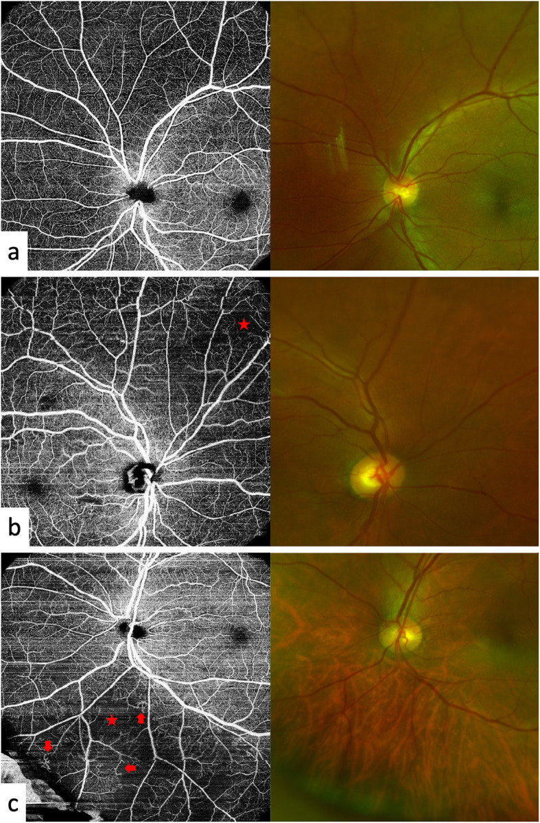
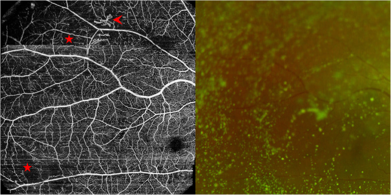
Prospective cross-sectional study. Fifty-five eyes of 30 diabetic patients without clinical retinal signs were included. All subjects underwent OCTA examination with a 12 × 12 mm field of view of 5 visual fixations (1 central fixation and 4 peripheral fixations) to compose a UWF OCTA image. In the UWF images, the central area corresponded to the original central image obtained using central fixation, and the peripheral area was the remaining area. Lesions, including nonperfusion areas (NPAs), microvascular dilation and tortuosity, and neovascularization (NV), were recorded in different areas. Diabetes history was also recorded.
Peripheral areas presented significantly more microvascular dilation and tortuosity than central areas (P = 0.024) and more NPAs than central areas, with borderline significance (P = 0.085). The number of lesion types was associated with HbA1c levels in the peripheral and overall areas (all P values < 0.001).
UWF SS OCTA is a promising imaging method for detecting vascular alterations in diabetic eyes without clinical signs to reveal retinal microvascular alterations. These alterations were correlated with systemic conditions.
使用超广角扫频光学相干断层扫描血管造影(UWF SS OCTA)研究临床前期糖尿病视网膜病变(DR)患者眼部视网膜微血管的变化。
前瞻性横断面研究。纳入30例无临床视网膜体征的糖尿病患者的55只眼。所有受试者均接受OCTA检查,采用12×12mm视野,5个视觉注视点(1个中央注视点和4个周边注视点)以组成UWF OCTA图像。在UWF图像中,中央区域对应于使用中央注视点获得的原始中央图像,周边区域为其余区域。记录不同区域的病变,包括无灌注区(NPA)、微血管扩张和迂曲以及新生血管(NV)。还记录了糖尿病病史。
周边区域的微血管扩张和迂曲明显多于中央区域(P = 0.024),无灌注区也多于中央区域,具有临界显著性(P = 0.085)。病变类型的数量与周边和整个区域的糖化血红蛋白水平相关(所有P值<0.001)。
UWF SS OCTA是一种很有前景的成像方法,可用于检测无临床体征的糖尿病患者眼部的血管变化,以揭示视网膜微血管变化。这些变化与全身状况相关。