Department of Biosciences, University of Salzburg, Vascular and Exercise Biology Research Group, Salzburg, Austria.
Department of Biomedicine, University of Basel, Basel, Switzerland.
Anat Rec (Hoboken). 2022 Feb;305(2):243-253. doi: 10.1002/ar.24649. Epub 2021 May 24.
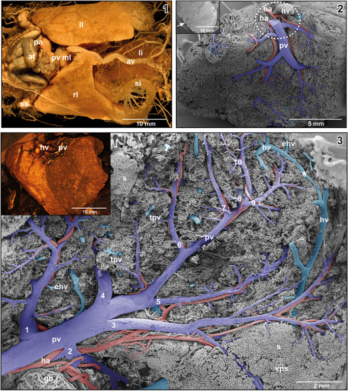
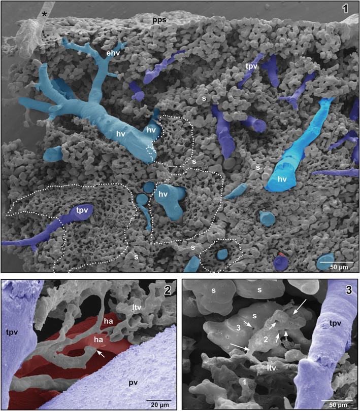
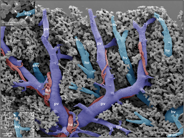
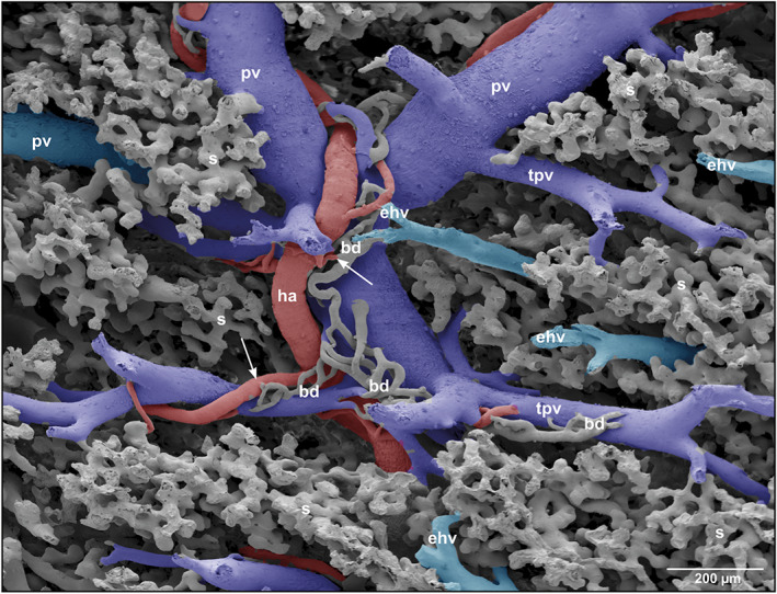
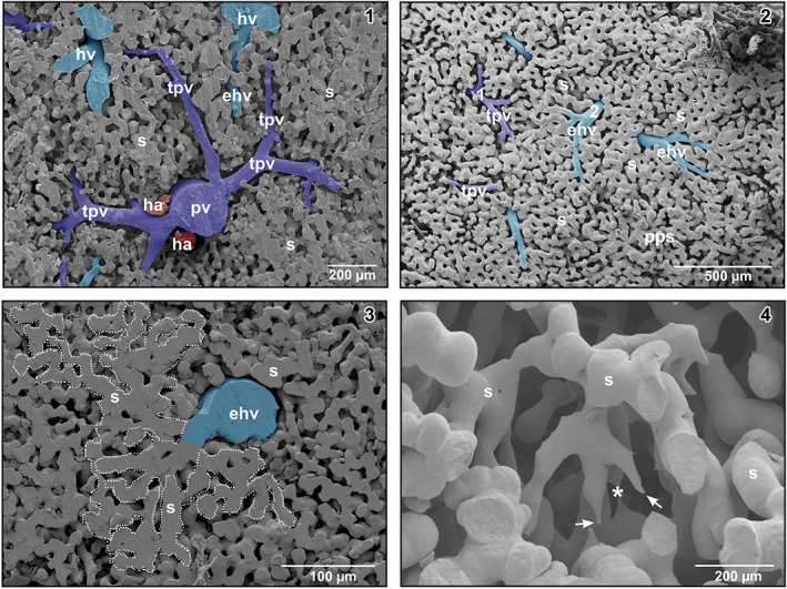
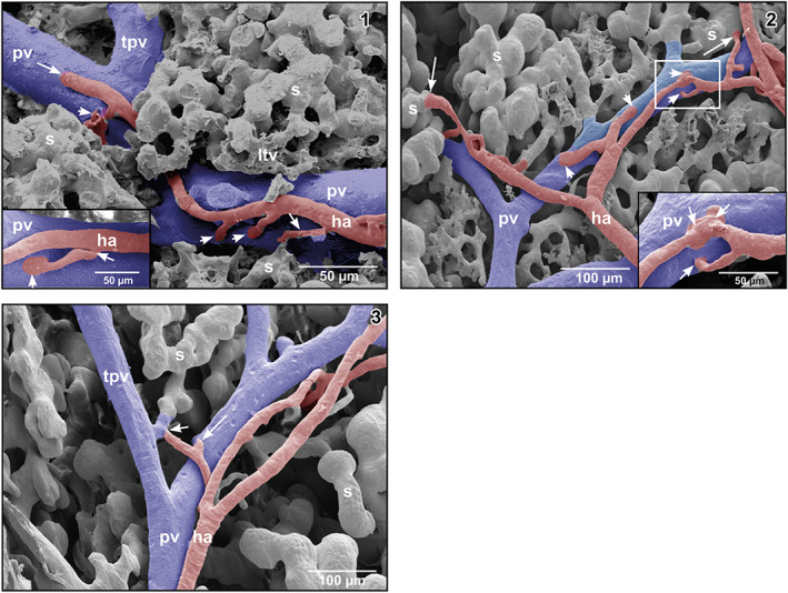
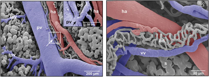
The microvascular anatomy of the non-lobulated liver of adult Xenopus laevis was studied by scanning electron microscopy of vascular corrosion casts. Hepatic portal veins and hepatic arteries entered hepatic lobes at the hiluses, hepatic veins left at these sites. Intraparenchymal, hepatic portal veins branched up to 10 times before terminal portal venules supplied liver sinusoids. Hepatic arteries closely followed portal vessels. Arteriolar side branches formed anastomoses with close by portal venules (arteriolar-portal anastomoses; APAs), liver sinusoids (arteriolar-sinusoidal anastomoses; ASAs), and peribiliary plexus vessels. Distally, hepatic arteries anastomosed with terminal portal venules having >100 μm in diameter. Liver sinusoids formed a dense three-dimensional network displaying signs of non-sprouting and sprouting angiogenesis evidenced by "holes" and blind ending tapering cast vascular structures (sprouts), respectively. Sinusoids drained via efferent hepatic veins. Right and left hepatic veins drained into the posterior caval vein. Locally, a dense honeycomb-like 3D-meshwork of resin structures was found around terminal portal venules and hepatic arteries. These networks were fed by hepatic arterioles and drained into adjacent terminal portal venules. As their morphologies differed significantly from sinusoids and they were found at sites where diffuse lymphoid tissue is described, we are convinced that they represent the vasculature of diffuse lymphoid tissue areas. Frequencies and diameter ratios of hepatic portal venules versus hepatic arterioles anastomosing with the former (APAs) implicate that the arterial supply contributes to the oxygenation of parenchymal and stromal cells rather than to a significant increase in blood flow towards hepatic sinusoids.
利用血管腐蚀铸型的扫描电子显微镜研究了成年非洲爪蟾非叶状肝脏的微血管解剖结构。肝门静脉和肝动脉在肝门处进入肝叶,肝静脉在此处离开。肝内门静脉分支多达 10 次,然后终末门静脉小静脉供应肝窦。肝动脉紧密跟随门静脉。小动脉侧支与附近的门静脉小静脉(动脉-门脉吻合;APAs)、肝窦(动脉-窦状隙吻合;ASAs)和胆管丛血管形成吻合。在远端,肝动脉与直径大于 100μm 的终末门静脉小静脉吻合。肝窦形成密集的三维网络,显示出非发芽和发芽血管生成的迹象,分别由“孔”和盲端逐渐变细的铸型血管结构(芽)证实。窦汇通过流出性肝静脉排出。右和左肝静脉汇入后腔静脉。局部可见,在终末门静脉小静脉和肝动脉周围存在密集的蜂窝状 3D 树脂结构网络。这些网络由肝动脉小动脉供应,并排入相邻的终末门静脉小静脉。由于它们的形态与窦明显不同,并且在描述弥漫性淋巴组织的部位发现,我们确信它们代表了弥漫性淋巴组织区域的血管结构。与前者吻合的肝门静脉小静脉与肝动脉小动脉的频率和直径比(APAs)表明,动脉供应有助于实质和基质细胞的氧合,而不是显著增加向肝窦的血流。