Cardobi Nicolò, Dal Palù Alessandro, Pedrini Federica, Beleù Alessandro, Nocini Riccardo, De Robertis Riccardo, Ruzzenente Andrea, Salvia Roberto, Montemezzi Stefania, D'Onofrio Mirko
Radiology Unit, Department of Pathology and Diagnostics, University Hospital of Verona, Piazzale Aristide Stefani, 1, 37126 Verona, Italy.
Department of Mathematical, Physical and Computer Sciences, University of Parma, 43121 Parma, Italy.
Cancers (Basel). 2021 Apr 30;13(9):2162. doi: 10.3390/cancers13092162.
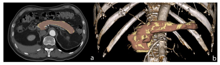
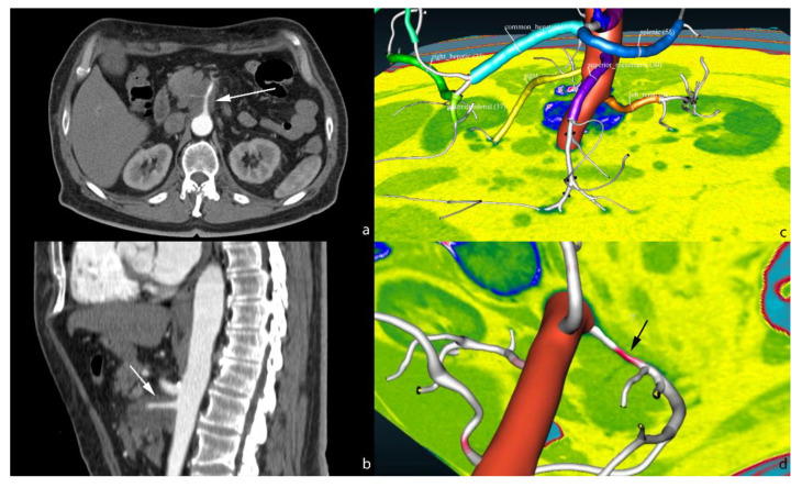
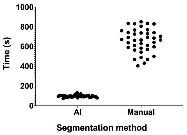
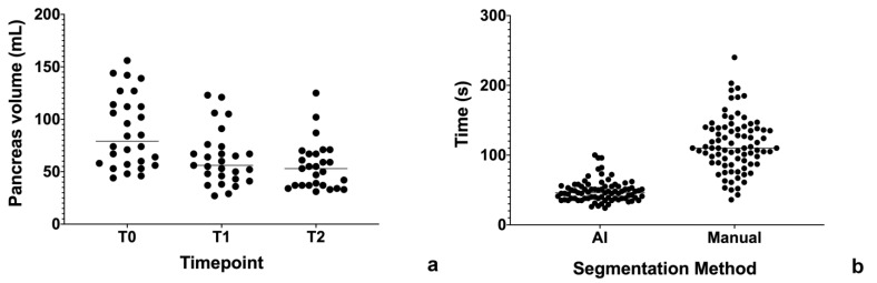
Artificial intelligence (AI) is one of the most promising fields of research in medical imaging so far. By means of specific algorithms, it can be used to help radiologists in their routine workflow. There are several papers that describe AI approaches to solve different problems in liver and pancreatic imaging. These problems may be summarized in four different categories: segmentation, quantification, characterization and image quality improvement. Segmentation is usually the first step of successive elaborations. If done manually, it is a time-consuming process. Therefore, the semi-automatic and automatic creation of a liver or a pancreatic mask may save time for other evaluations, such as quantification of various parameters, from organs volume to their textural features. The alterations of normal liver and pancreas structure may give a clue to the presence of a diffuse or focal pathology. AI can be trained to recognize these alterations and propose a diagnosis, which may then be confirmed or not by radiologists. Finally, AI may be applied in medical image reconstruction in order to increase image quality, decrease dose administration (referring to computed tomography) and reduce scan times. In this article, we report the state of the art of AI applications in these four main categories.
人工智能(AI)是迄今为止医学成像领域最具前景的研究领域之一。通过特定算法,它可用于协助放射科医生进行日常工作流程。有几篇论文描述了人工智能解决肝脏和胰腺成像中不同问题的方法。这些问题可归纳为四个不同类别:分割、量化、特征描述和图像质量改善。分割通常是后续详细分析的第一步。如果手动进行,这是一个耗时的过程。因此,半自动和自动创建肝脏或胰腺掩码可为其他评估节省时间,例如从器官体积到其纹理特征的各种参数的量化。正常肝脏和胰腺结构的改变可能提示弥漫性或局灶性病变的存在。可以训练人工智能识别这些改变并提出诊断,然后由放射科医生确认或否定。最后,人工智能可应用于医学图像重建,以提高图像质量、减少剂量(指计算机断层扫描)并缩短扫描时间。在本文中,我们报告了人工智能在这四个主要类别中的应用现状。